
Fig. 2 - Supplemental 3
- ID
- ZDB-FIG-250424-33
- Publication
- Scerbo et al., 2025 - In vivo targeted and deterministic single-cell malignant transformation
- Other Figures
- All Figure Page
- Back to All Figure Page
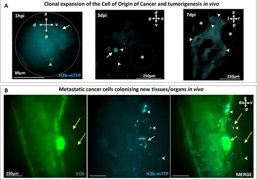
Tracking of the clonal expansion of a photoinduced cell in one embryo over 7 days. (A) At 1 day post-fertilization (dpf), a single cell (white arrowhead) in the vicinity of the otic vesicle (white arrow) was photo-induced to express the oncogene KRASG12V, identified within ~1 hr (1 hr post-induction [hpi]) by the fluorescent H2B-mTFP marker. Transient (24 hr) dexamethasone (DEX) activation of Ventx-GR was done following photoactivation resulting in malignant transformation of the induced cell and clonal expansion. At 3 day post-induction (dpi) a few cells (white arrowheads) progeny of the induced one are observed in the vicinity of the otic vesicle (white arrow). At 7 dpi, a tumor mass is observed in the brain (white arrowheads) and some of the cells (B) have metastasized (white arrowheads) to colonize new tissues as the proctodeum, close to the ventral fin (yellow arrows). Body axes are shown (a: anterior; p: posterior; d: dorsal; v: ventral; l: left; r: right). |