
Fig. 1
- ID
- ZDB-FIG-250401-30
- Publication
- Korenkova et al., 2024 - Tunneling nanotubes enable intercellular transfer in zebrafish embryos
- Other Figures
- All Figure Page
- Back to All Figure Page
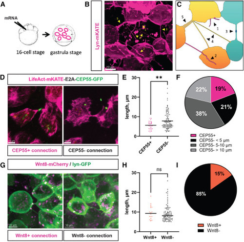
Characterization of intercellular connections in zebrafish gastrula (A) Schematic representation of the labeling strategy to visualize intercellular connections: zebrafish embryo is injected into one of the central cells at the 16-cell stage, incubated at 28°C until it reaches the gastrula stage (7–9 hpf), and mounted for imaging. (B) Fluorescent photograph of different protrusions that can be observed at the gastrula stage following membrane labeling by lyn-mKATE mRNA injection, identified by yellow arrowheads: (1) short protrusions, (2) long closed-ended protrusions, (3) connections with interrupted membrane labeling, (4) thin TNT-like structures, and (5) thick TNT-like structures. (C) Cartoon representing the image (B), suggesting the coexistence of (1) filopodia, (2) cytonemes, (3) cytokinetic bridges, (4) TNT-like structures, and (5) bundles of TNT-like structures. (D) Representative images of CEP55-positive cytokinetic bridges and CEP55-negative TNT-like connections. (E) Length distribution and average lengths between CEP55-positive (n = 27 connections, mean 5.6 μm) and CEP55-negative connections (n = 114 connections, mean 7.8 μm), p = 0.0096, statistical analysis is t test. (F) Relative numbers of CEP55-positive connections and CEP55-negative TNT-like connections having lengths below 5 μm, from 5 to 10 μm, and above 10 μm (n = 141 connections). (G) Representative images of Wnt8a-bearing cytonemes and Wnt8a-negative TNT-like connections. (H) Length distribution and average lengths of Wnt8a-positive (n = 18 connections, mean 9.7 μm) and Wnt8a-negative connections (n = 100 connections, mean 9.4 μm), p = 0.7228, statistical analysis is t test. (I) Relative numbers of Wnt8a-positive cytonemes and Wnt8a-negative TNT-like connections (n = 118 connections). Scale bars are 10 μm. |
Reprinted from Developmental Cell, 60, Korenkova, O., Liu, S., Prlesi, I., Pepe, A., Albadri, S., Del Bene, F., Zurzolo, C., Tunneling nanotubes enable intercellular transfer in zebrafish embryos, 524-534.e3, Copyright (2024) with permission from Elsevier. Full text @ Dev. Cell