|
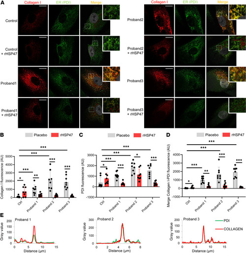
Treatment with rHSP47 reduces intracellular procollagen retention. (A–D) Representative images (A) and quantification of immunofluorescence of (B) collagen I (aligned rank ANOVA [ARA] test, FARA = 18.06, P < 0.001 by overall test), (C) ER marker protein disulfide isomerase (PDI) (ARA test, FARA = 15.12, P < 0.001 by overall test), and of (D) collagen I–PDI signal (ARA test, FARA = 40.35, P < 0.001 by overall test) of osteogenesis imperfecta (OI) proband and control fibroblasts treated for 16 hours with 0.5 μM rHSP47 or with placebo. Asterisks in B–D indicate the post hoc test results. Biological replicates (n = 3) were performed. For each biological replicate, the signal was quantified on 8 images (original magnification, ×40) for each genotype/condition (number of cells, >90). Error bars indicate SD. (E) The colocalization of collagen I–PDI signal in proband 1, 2, and 3 cells is evident by the 2 overlapping peaks in the graph (n = 3). Nuclei were counterstained with DAPI. Scale bars: 15 μm. Zoomed inset magnification, ×3. Statistical analyses details are reported in Supplemental Table 2. *P < 0.05; **P ≤ 0.01; ***P ≤ 0.001.
|