
FIGURE
Fig. 3
- ID
- ZDB-FIG-250324-24
- Publication
- Qian et al., 2025 - Toxic effects of prolonged propofol exposure on cardiac development in zebrafish larvae
- Other Figures
- All Figure Page
- Back to All Figure Page
Fig. 3
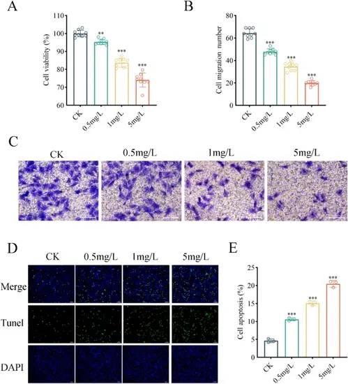
Propofol inhibits H9C2 cardiomyocyte viability, migration, and induces apoptosis. Propofol inhibited cell viability (A) and migration (B) in a concentration-dependent manner. Representative images from Transwell assays (C) illustrate its impact on cell migration, and Tunel assays (D) demonstrate propofol-induced apoptosis, which increased with higher concentrations (E). Data shown as the means ± SD, (n = 3). Scale bars = 50 μm. **p < 0.01, ***p < 0.001 vs. control |
Expression Data
Expression Detail
Antibody Labeling
Phenotype Data
Phenotype Detail
Acknowledgments
This image is the copyrighted work of the attributed author or publisher, and
ZFIN has permission only to display this image to its users.
Additional permissions should be obtained from the applicable author or publisher of the image.
Full text @ BMC Anesthesiol