
Fig. 2
- ID
- ZDB-FIG-250324-23
- Publication
- Qian et al., 2025 - Toxic effects of prolonged propofol exposure on cardiac development in zebrafish larvae
- Other Figures
- All Figure Page
- Back to All Figure Page
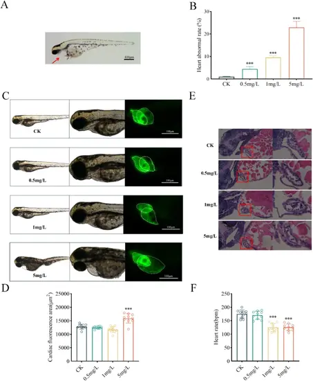
Propofol alters heart structure and function (heart rate) in zebrafish larvae. (A). Propofol-induced pericardial cyst in zebrafish larvae at 96hpf. (B). The incidence of propofol-induced heart malformations rose with higher concentrations. Cardiac malformations are defined as the observation of pericardial edema, abnormal chamber size, pericardial extension, and irregular heart morphology. (C). Cardiac morphology changes in Tg [myl7:eGFP] zebrafish larvae exposed to varying propofol concentrations included reduced atrioventricular overlap, prolonged pericardium, and enlarged cardiac cavity area. (D). Cardiac fluorescence area. (E). Representative images of HE-stained zebrafish embryo hearts following propofol exposure. (F). Heart rate changes in zebrafish larvae treated with different concentrations of propofol. Data are presented as the means ± SD of at least three independent experiments. Student’s t-test statistical analysis. ***p < 0.001 vs. control |