
Fig. 6
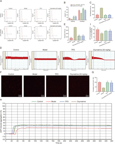
Oxymatrine accelerates the recovery of platelet function in irradiated mice. (A) Flow cytometry analysis of CD62P+ cells in washed platelets in control, model, thrombopoietin (TPO)-treated, and oxymatrine-treated groups (top), and the activated (adenosine diphosphate (ADP) 10 μM) CD62P+ cells in each group (bottom). (B) The histogram shows the percentage of CD62P+cells with or without ADP (10 μM) activation (n = 3 per group). (C) Tail bleeding time of each group after treatment of 12 days (n = 3 per group). (D) Analysis of carotid blood flow detection of each group. (E) The mean carotid artery occlusion times are represented by the histogram (n = 3 per group). (F) Micrographs of collagen-coated slides with the same number of platelets perfused. Red represents platelets. (G) The histogram shows the average coverage of red fluorescence on the whole surface by ImageJ software (n = 3 per group). (H) Platelet aggregation was detected under the activation of ADP by the device of turbidimetric aggregation-monitoring. (I) The histogram showed the maximum aggregation amplitude of platelets in each group (n = 3 per group). Data represent the mean ± standard deviation (SD) of three independent experiments. ∗P < 0.05, ∗∗P < 0.01, and ∗∗∗P < 0.001 vs. the model group. |