
Fig. 4
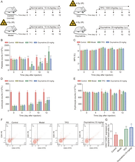
Oxymatrine accelerates platelet recovery in acute X-ray irradiated mice. (A) The strategies of establishing the irradiated mice and treatment of each groups. (B) Peripheral platelet level of control group, thrombopoietin (TPO)-treated group (3000 U/kg), oxymatrine-treated group (20 mg/kg), and model group. Histology analysis was performed on days 0, 4, 7, 10, and 12 (n = 6 per group). (C) Mean platelet volume (MPV) values in each groups (n = 6 per group). (D) Leukocyte counts in each group (n = 6 per group). (E) Erythrocyte counts in each group (n = 6 per group). (F) Flow cytometry analysis of expression of CD41 and CD61 in peripheral blood (PB) platelets in each group after treatment for 12 days. (G) The histogram exhibits the percentage of expression of CD41+/CD61+ in PB cells in each group (n = 3 per group). Data represent the mean ± standard deviation (SD). Two-way analysis of variance (ANOVA) with Tukey's multiple comparisons test was used unless otherwise specified. ∗P < 0.05, ∗∗P < 0.01, and ∗∗∗P < 0.001 vs. the corresponding model group. IR: ionizing radiation; FITC: fluorescein isothiocyanate; UL: upper left; UR: upper right; LL: lower left; LR: lower right. |