
Fig. 1
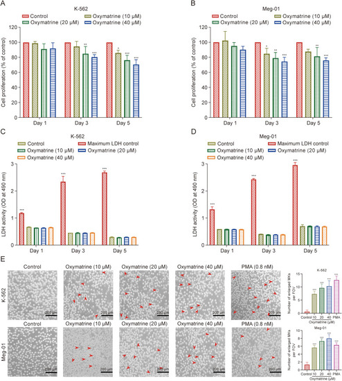
Screening safe concentrations of oxymatrine for the treatment of K-562 and Meg-01 megakaryocyte (MK). (A, B) The Cell Counting Kit-8 (CCK-8) assay for oxymatrine-treated K-562 (A) and Meg-01 (B) MK proliferation. Three different time points and drug concentrations were used to measure the cell proliferation rate (n = 6 per group). (C, D) Detection of lactate dehydrogenase (LDH) release of K-562 (C) and Meg-01 (D) cells treated with different drug concentrations and measured at different time points (n = 6 per group). (E) Representative images of oxymatrine intervened K-562 and Meg-01 cells with various concentration (10, 20, and 40 μM) for five days (n = 3 per group). Phorbol 12-myristate 13-acetate (PMA) was used as positive control (0.8 nM). Data represent the mean ± standard deviation (SD) of six or three independent experiments and are analyzed by one-way analysis of variance (ANOVA) with Dunnett's 154. ∗P < 0.05, ∗∗P < 0.01, and ∗∗∗P < 0.001 vs. the control. FOV: field of view. |