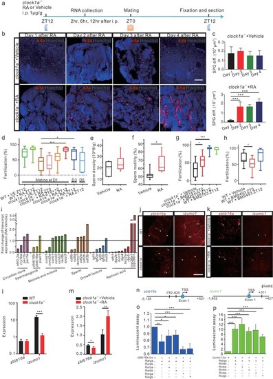
atRA treatment rescues the clock1a−/− spermatogenesis and fertilization defects via zbtb16a and izumo1 in zebrafish. (a) Schedules of the experiments. (b) FISH images of the WT and clock1a−/− testis sections with the kita probe at days 1 to 4 following the atRA or vehicle treatment i.p. at ZT12 (n = 3 × 5). The signals were superimposed with the Hoechst33423 nuclear counterstain. Scale bar, 100 μm. (c) Quantification of kita-positive cells in b (n = 4). (d) Fertilization rates by pairwise crosses of WT males or clock1a−/− males with WT females 1, 3, and 5 days after atRA or vehicle treatment at ZT0 or ZT12 (n = 9–18). (e and f) Sperm density (e) and motility (f) of the clock1a−/− mutant males after atRA or vehicle treatment (n = 6). (g and h) Fertilization rates by pairwise crosses of WT females with clock1a−/− males or WT males treated with BMS753 (g), and WT females with WT males treated with BMS493 (h) at ZT0 or ZT12 (n = 10–18). (i) The ratios of gene expression levels in the clock1a−/− testis 2 hours after atRA treatment to those after vehicle treatment (n = 3). (j) FISH images of WT and clock1a−/− testes using zbtb16a or izumo1 probe (n = 4). Scale bar, 100 μm. (k) FISH images of clock1a−/− testes treated with RA or vehicle using zbtb16a or izumo1 probe (n = 4). (l and m) Quantification of zbtb16a-positive and izumo1-positive cells in (j and k) (n = 4). (n) Schematic diagrams of the RARE motifs-containing izumo1 and zbtb16a promoters. (o and p) Luciferase reporter assays of izumo1 (o) and zbtb16a (p) promoters (n = 3). (b, j and k) Arrows indicate spermatogonia. Sc, spermatocyte; Sz, spermatozoon. *P < 0.05; **P < 0.01;***P < 0.001 (Fig. S5).
|
