
Fig. 3
- ID
- ZDB-FIG-250220-14
- Publication
- Kim et al., 2024 - CRISPR-Cas13d as a molecular tool to achieve targeted gene expression knockdown in chick embryos
- Other Figures
- All Figure Page
- Back to All Figure Page
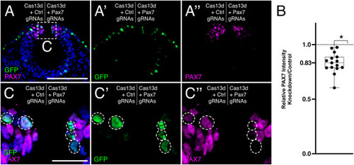
One-color CRISPR-Cas13d-mediated PAX7 knockdown. (A) A representative transverse cross-section of a HH9 chick embryo head bilaterally electroporated with Cas13d + Control guide RNA (gRNA) plasmids (left), and Cas13d + Pax7 gRNA plasmids (right). Reduced PAX7 intensity is observed on the right side of the embryo. Scale bar, 100 μm. (B) Quantification of PAX7 knockdown. Each data point represents the relative PAX7 intensity detected on the right side of an embryo (PAX7 knockdown) compared to the left side of the same embryo (contralateral control). The relative intensity measurements for each embryo are averages of the intensities detected from three nonadjacent cross-sections. A statistically significant reduction in PAX7 intensity is observed (83.1 ± 2.4% of the contralateral control side; n = 15 embryos) with CRISPR-Cas13d-mediated knockdown. ∗, p < 0.0001; one-sample Wilcoxon signed-rank test. (C) Enlarged view of boxed area shown in (A). GFP+ cells are highlighted by white circles. Only the GFP+ cells (C′) on the right side of the embryo show reduction in PAX7 intensity (C″). Scale bar, 20 μm. (For interpretation of the references to color in this figure legend, the reader is referred to the Web version of this article.) |
Reprinted from Developmental Biology, , Kim, M., Hutchins, E.J., CRISPR-Cas13d as a molecular tool to achieve targeted gene expression knockdown in chick embryos, , Copyright (2024) with permission from Elsevier. Full text @ Dev. Biol.