
Fig. 1
- ID
- ZDB-FIG-250220-12
- Publication
- Kim et al., 2024 - CRISPR-Cas13d as a molecular tool to achieve targeted gene expression knockdown in chick embryos
- Other Figures
- All Figure Page
- Back to All Figure Page
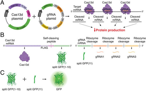
Delivery strategy of a two-plasmid CRISPR-Cas13d system for avian embryos. (A) Schematic depicting structure of Cas13d and guide RNA (gRNA) plasmids and the in vivo knockdown effect. Plasmid expression is driven by a ubiquitous promoter (CAG). Cas13d protein and gRNAs form ribonucleoprotein complexes to cleave target mRNA at multiple sites across the coding region, ultimately resulting in disrupted translation and decreased protein production. (B) The Cas13d plasmid generates a FLAG-tagged Cas13d protein and a split GFP(1–10) non-fluorescent reporter protein, separated by a T2A self-cleaving peptide. The gRNA plasmid generates a split GFP(11) non-fluorescent reporter protein, and three unique gRNAs flanked by ribozyme sequences (HH and HDV) that separate the transcribed components. (C) When co-expressed, the split GFP non-fluorescent reporters GFP(1–10) and GFP(11) self-complement into a functional, fluorescent GFP reporter protein to label cells that have successfully received both plasmids. HH, hammerhead ribozyme sequence; HDV, hepatitis delta virus ribozyme sequence. |
Reprinted from Developmental Biology, , Kim, M., Hutchins, E.J., CRISPR-Cas13d as a molecular tool to achieve targeted gene expression knockdown in chick embryos, , Copyright (2024) with permission from Elsevier. Full text @ Dev. Biol.