
Fig. 3
- ID
- ZDB-FIG-250218-78
- Publication
- Wang et al., 2025 - Direct lysine dimethylation of IRF3 by the methyltransferase SMYD3 attenuates antiviral innate immunity
- Other Figures
- All Figure Page
- Back to All Figure Page
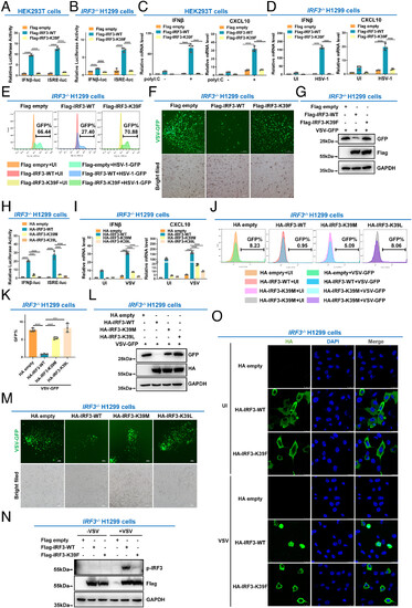
IRF3 is inactivated by methylation at lysine 39. (A) Luciferase activity of IFNβ promoter reporter and ISRE reporter in HEK293T cells transfected with empty Flag vector, WT IRF3 (Flag-IRF3-WT), or the methylation mimic mutant (Flag-IRF3-K39F). (B) Luciferase activity of the IFN-β promoter reporter and ISRE reporter in IRF3−/− H1299 cells transfected with empty Flag vector, WT IRF3 (Flag-IRF3-WT), or the methylation mimic mutant (Flag-IRF3-K39F). (C) qPCR analysis of IFNβ and CXCL10 mRNA in HEK293T cells transfected with empty Flag vector, WT IRF3 (Flag-IRF3-WT), or the methylation mimic mutant (Flag-IRF3-K39F), followed by transfection with (+) or without (−) poly I: C. (D) qPCR analysis of IFNβ and CXCL10 mRNA in IRF3−/− H1299 cells transfected with empty Flag vector, WT IRF3 (Flag-IRF3-WT), or the methylation mimic mutant (Flag-IRF3-K39F), followed by UI or infected with HSV-1. (E) Flow cytometric analysis of HSV-1-GFP virus replication in IRF3−/− H1299 cells transfected with empty Flag vector, WT IRF3 (Flag-IRF3-WT), or the methylation mimic mutant (Flag-IRF3-K39F), followed by UI or infected with HSV-1-GFP virus. (F) Microscopic imaging of VSV-GFP virus replication in IRF3−/− H1299 cells transfected with empty Flag vector, WT IRF3 (Flag-IRF3-WT), or the methylation mimic mutant (Flag-IRF3-K39F), followed by infection with VSV-GFP virus. (G) Immunoblotting analysis of VSV-GFP virus replication in IRF3−/− H1299 cells transfected with empty Flag vector, WT IRF3 (Flag-IRF3-WT), or the methylation mimic mutant (Flag-IRF3-K39F), followed by infection with VSV-GFP virus. (H) Luciferase activity of the IFN-β promoter reporter and ISRE reporter in IRF3−/− H1299 cells transfected with empty HA vector, WT IRF3 (HA-IRF3-WT), or the methylation mimic mutant (HA-IRF3-K39M or HA-IRF3-K39L mutant). (I) qPCR analysis of IFNβ and CXCL10 mRNA in IRF3−/− H1299 cells transfected with empty HA vector, WT IRF3 (HA-IRF3-WT), or the methylation mimic mutant (HA-IRF3-K39M or HA-IRF3-K39L mutant), followed by UI or infected with VSV. (J and K) Flow cytometric analysis of VSV-GFP virus replication in IRF3−/− H1299 cells transfected with empty HA vector, WT IRF3 (HA-IRF3-WT), or the methylation mimic mutant (HA-IRF3-K39M or HA-IRF3-K39L mutant), followed by UI or infected with VSV-GFP virus. (L) Immunoblotting analysis of VSV-GFP virus replication in IRF3−/− H1299 cells transfected with empty HA vector, WT IRF3 (HA-IRF3-WT), or the methylation mimic mutant (HA-IRF3-K39M or HA-IRF3-K39L mutant), followed by infection with VSV-GFP virus. (M) Microscopic imaging of VSV-GFP virus replication in IRF3−/− H1299 cells transfected with empty HA vector, WT IRF3 (HA-IRF3-WT), or the methylation mimic mutant (HA-IRF3-K39M or HA-IRF3-K39L mutant), followed by infection with VSV-GFP virus. (N) Phosphorylation of IRF3 in IRF3−/− H1299 cells transfected with empty Flag vector, WT IRF3 (Flag-IRF3-WT), or the methylation mimic mutant (Flag-IRF3-K39F), followed by VSV infection for 8 h.(O) Localization of WT IRF3 (HA-IRF3-WT) and the methylation mimic mutant (HA-IRF3-K39F) in IRF3−/− H1299 cells UI or infected with VSV. ****P < 0.0001. Graphs represent fold induction relative to the untreated cells. Data are presented as the mean values of a representative experiment performed in triplicate (A–D, H, I, and K) or as representative data (E–G, J, and L–O); these experiments were repeated independently at least three times, and error bars indicate S.D. |