
|
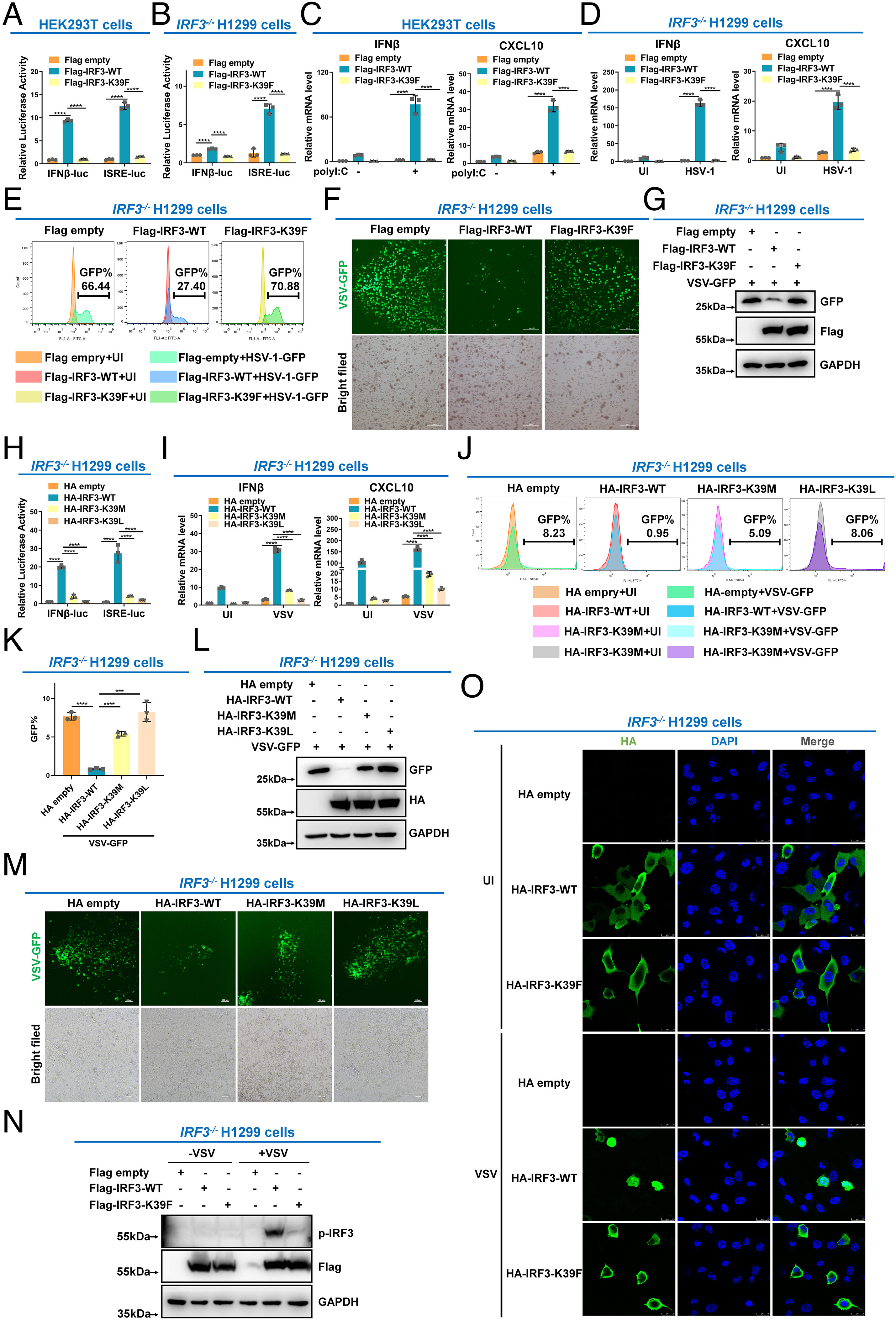
Fig. 3 IRF3 is inactivated by methylation at lysine 39. (A) Luciferase activity of IFNβ promoter reporter and ISRE reporter in HEK293T cells transfected with empty Flag vector, WT IRF3 (Flag-IRF3-WT), or the methylation mimic mutant (Flag-IRF3-K39F). (B) Luciferase activity of the IFN-β promoter reporter and ISRE reporter in IRF3−/− H1299 cells transfected with empty Flag vector, WT IRF3 (Flag-IRF3-WT), or the methylation mimic mutant (Flag-IRF3-K39F). (C) qPCR analysis of IFNβ and CXCL10 mRNA in HEK293T cells transfected with empty Flag vector, WT IRF3 (Flag-IRF3-WT), or the methylation mimic mutant (Flag-IRF3-K39F), followed by transfection with (+) or without (−) poly I: C. (D) qPCR analysis of IFNβ and CXCL10 mRNA in IRF3−/− H1299 cells transfected with empty Flag vector, WT IRF3 (Flag-IRF3-WT), or the methylation mimic mutant (Flag-IRF3-K39F), followed by UI or infected with HSV-1. (E) Flow cytometric analysis of HSV-1-GFP virus replication in IRF3−/− H1299 cells transfected with empty Flag vector, WT IRF3 (Flag-IRF3-WT), or the methylation mimic mutant (Flag-IRF3-K39F), followed by UI or infected with HSV-1-GFP virus. (F) Microscopic imaging of VSV-GFP virus replication in IRF3−/− H1299 cells transfected with empty Flag vector, WT IRF3 (Flag-IRF3-WT), or the methylation mimic mutant (Flag-IRF3-K39F), followed by infection with VSV-GFP virus. (G) Immunoblotting analysis of VSV-GFP virus replication in IRF3−/− H1299 cells transfected with empty Flag vector, WT IRF3 (Flag-IRF3-WT), or the methylation mimic mutant (Flag-IRF3-K39F), followed by infection with VSV-GFP virus. (H) Luciferase activity of the IFN-β promoter reporter and ISRE reporter in IRF3−/− H1299 cells transfected with empty HA vector, WT IRF3 (HA-IRF3-WT), or the methylation mimic mutant (HA-IRF3-K39M or HA-IRF3-K39L mutant). (I) qPCR analysis of IFNβ and CXCL10 mRNA in IRF3−/− H1299 cells transfected with empty HA vector, WT IRF3 (HA-IRF3-WT), or the methylation mimic mutant (HA-IRF3-K39M or HA-IRF3-K39L mutant), followed by UI or infected with VSV. (J and K) Flow cytometric analysis of VSV-GFP virus replication in IRF3−/− H1299 cells transfected with empty HA vector, WT IRF3 (HA-IRF3-WT), or the methylation mimic mutant (HA-IRF3-K39M or HA-IRF3-K39L mutant), followed by UI or infected with VSV-GFP virus. (L) Immunoblotting analysis of VSV-GFP virus replication in IRF3−/− H1299 cells transfected with empty HA vector, WT IRF3 (HA-IRF3-WT), or the methylation mimic mutant (HA-IRF3-K39M or HA-IRF3-K39L mutant), followed by infection with VSV-GFP virus. (M) Microscopic imaging of VSV-GFP virus replication in IRF3−/− H1299 cells transfected with empty HA vector, WT IRF3 (HA-IRF3-WT), or the methylation mimic mutant (HA-IRF3-K39M or HA-IRF3-K39L mutant), followed by infection with VSV-GFP virus. (N) Phosphorylation of IRF3 in IRF3−/− H1299 cells transfected with empty Flag vector, WT IRF3 (Flag-IRF3-WT), or the methylation mimic mutant (Flag-IRF3-K39F), followed by VSV infection for 8 h.(O) Localization of WT IRF3 (HA-IRF3-WT) and the methylation mimic mutant (HA-IRF3-K39F) in IRF3−/− H1299 cells UI or infected with VSV. ****P < 0.0001. Graphs represent fold induction relative to the untreated cells. Data are presented as the mean values of a representative experiment performed in triplicate (A–D, H, I, and K) or as representative data (E–G, J, and L–O); these experiments were repeated independently at least three times, and error bars indicate S.D.