
Fig. 2
- ID
- ZDB-FIG-250218-77
- Publication
- Wang et al., 2025 - Direct lysine dimethylation of IRF3 by the methyltransferase SMYD3 attenuates antiviral innate immunity
- Other Figures
- All Figure Page
- Back to All Figure Page
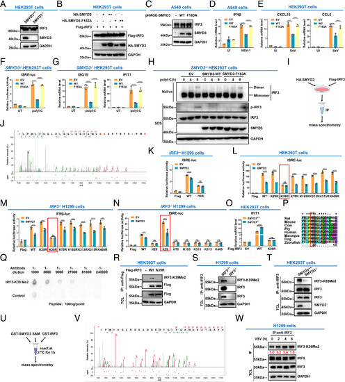
SMYD3 catalyzes the methylation of IRF3 at lysine 39. (A) Immunoblotting of endogenous IRF3 in SMYD3+/+ or SMYD3−/− HEK293T cells. (B) Immunoblotting of ectopic IRF3 protein in HEK293T cells transfected with WT SMYD3 or the enzymatically inactive mutant of SMYD3 (F183A) at increasing doses. (C) Immunoblotting of endogenous IRF3 in A549 cells infected with pHAGE-SMYD3, or pHAGE-SMYD3(F183A) lentiviruses. (D) qPCR analysis of IFNβ in A549 cells transfected with empty vector (EV), WT SMYD3 (WT), or the enzymatically inactive mutant of SMYD3 (F183A), followed by UI or infected with HSV-1 for 8 h. (E) qPCR analysis of CXCL10 and CCL5 in HEK293T cells transfected with empty vector (EV), WT SMYD3 (WT), or the enzymatically inactive mutant of SMYD3 (F183A), followed by UI or infected with SeV for 8 h. (F) Luciferase activity of the ISRE reporter in SMYD3−/− HEK293T cells transfected with empty vector (EV), WT SMYD3 (WT), or the enzymatically inactive mutant of SMYD3 (F183A), followed by untransfected (UT) or transfected with poly I:C for 8 h. (G) qPCR analysis of ISG15 and IFIT1 in SMYD3−/− HEK293T cells transfected with empty vector (EV), WT SMYD3 (WT), or the enzymatically inactive mutant of SMYD3 (F183A), followed by untransfected (UT) or transfected with poly I:C for 8 h. (H) Phosphorylation and dimerization of IRF3 in SMYD3−/− HEK293T cells transfected with empty vector (EV), WT SMYD3 (WT), or the enzymatically inactive mutant of SMYD3 (F183A), followed by untransfected (UT) or transfected with poly I:C for 0, 4, and 8 h. (I) Scheme of detection of IRF3 methylation by mass spectrometry analysis. (J) Mass spectrometry analysis revealed that lysine 39 in IRF3 was dimethylated by SMYD3. (K) Luciferase activity of the ISRE reporter in IRF3−/− H1299 cells transfected with SMYD3 together with WT IRF3 (WT) or the lysine to arginine mutant (all the seven lysine residues, including K29, K39, K70, K193, K313, K315, and K409, were simultaneously mutated to arginine residues). (L) Luciferase activity of the ISRE reporter in HEK293T cells transfected with SMYD3 together with WT IRF3 (WT) or the lysine to arginine mutants, including K29R, K39R, K70R, K193R, K313R, K315R, or K409R. (M) Luciferase activity of the IFNβ promoter reporter in IRF3−/− H1299 cells transfected SMYD3 together with WT IRF3 (WT), or its lysine to arginine mutants. (N) Luciferase activity of the ISRE reporter in IRF3−/− H1299 cells transfected with SMYD3 together with WT IRF3 (WT), or its lysine to arginine mutants, including K29, K39, K70, K193, K313, K315, or K409 (In which only K29, K39, K70, K193, K313, K315, or K409, was kept intact respectively and other six lysine residues were simultaneously mutated to arginine residues). (O) qPCR analysis of IFIT1 in SMYD3+/+ and SMYD3−/− HEK293T cells transfected with empty vector (EV), WT IRF3 (WT), or IRF3-K39R mutant. (P) Sequence alignment of partial IRF3 (34-50 amino acids) from human, mouse, rat, cow, pig, macaque, dog, and zebrafish. The red box indicates a conserved lysine (K39). (Q) Dot blot assay for the specificity of the anti-IRF3-K39Me2 antibody. Equal amounts of dimethylated peptides or the control peptides were immunoblotted with the indicated dilutions of anti-IRF3-K39Me2 antibody. (R) HEK293T cells were transfected with empty Flag vector (−), Flag-IRF3 (WT), or Flag-IRF3-K39R, followed by immunoprecipitating with anti-Flag antibody, and immunoblotting with anti-IRF3-K39Me2 antibody. (S) Cell lysates from IRF3-intact or IRF3-null H1299 cells (IRF3+/+ or IRF3−/−) were extracted, followed by immunoprecipitation with anti-IRF3 antibody, and immunoblotting with anti-IRF3 and anti-IRF3-K39Me2 antibodies. (T) Cell lysates from SMYD3-intact or SMYD3-null HEK293T cells (SMYD3+/+ or SMYD3−/−) were extracted, followed by immunoprecipitation with anti-IRF3 antibody, and immunoblotting with anti-IRF3 and anti-IRF3-K39Me2 antibodies. (U) Scheme for the in vitro methylation assay and detection of IRF3 methylation by mass spectrometry analysis. (V) Mass spectrometry analysis revealed that lysine 39 in IRF3 was methylated by bacterially expressed SMYD3. (W) IRF3 methylation at lysine 39 was steadily increased after VSV infection. Anti-IRF3 antibody was used for Co-IP, and anti-IRF3-K39Me2 antibody was used for detection. IP, immunoprecipitation; TCL, total cell lysate; empty vector (EV); uninfected (UI); UT, untransfected; ns, not significant, *P < 0.05, **P < 0.01, ***P < 0.001, and ****P < 0.0001. Graphs represent fold induction relative to the untreated cells. Data are presented as the mean values of a representative experiment performed in triplicate (D–G and K–O) or as representative data (A–C, H, R–T, and W); these experiments were repeated independently at least three times, and error bars indicate S.D. |