
Fig. 4
- ID
- ZDB-FIG-250131-55
- Publication
- Chiba et al., 2024 - Zonated Wnt/β-catenin signal-activated cardiomyocytes at the atrioventricular canal promote coronary vessel formation in zebrafish
- Other Figures
- All Figure Page
- Back to All Figure Page
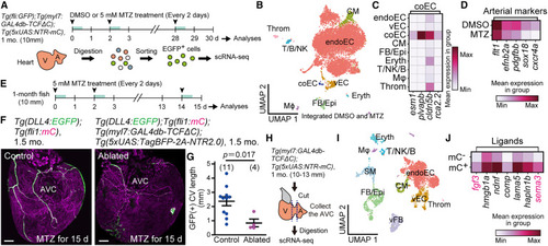
β-cat ON CMs are required for CV arterialization (A) An experimental design of preparation of cells followed by scRNA-seq. fli1:EGFP+ cells were collected from the fish treated with DMSO or 5 mM MTZ every 2 days. (B) Uniform manifold approximation and projection (UMAP) visualization of integrated cells from DMSO-treated and MTZ-treated hearts. Cells are color-coded by type. endoEC, endocardial EC; coEC, coronary EC; vEC, valvular EC; Epi, epicardial cells; FB, fibroblasts; Eryth, erythrocytes; T, T cells; NK, natural killer cells; B, B cells; Mϕ, macrophages; Throm, thrombocytes. (C) A matrix plot of coEC marker gene expression (bottom) in each cluster (left) shown in (B). (D) A matrix plot of arterial marker gene expression (bottom) of coECs from hearts treated with DMSO (upper) or MTZ (lower). (E) Experimental design to analyze the CV of the fish used in (F). Fish were treated with 5 mM MTZ every 2 days and analyzed after 15 days of treatment. (F) Representative images of arteries and CVs of control or Tg(DLL4:EGFP);Tg(myl7:GAL4db-TCFΔC);Tg(5xUAS:TagBFP-2A-NTR2.0) fish at 1.5 months treated with MTZ. A control heart (left) and a β-cat ON CM-ablated heart (right). (G) Quantitative analysis of the data shown in (F). (H) Experimental design for preparing the cells at the AVC followed by scRNA-seq. The fish indicated at the top treated with DMSO for 48 h were used. (I) UMAP visualization of cells obtained from the AVC of the fish in (H) at 1 month. Cells are color-coded by type. vFB, valvular FB; SM, smooth muscle cells. (J) A matrix plot of ligand expression (bottom) in β-cat OFF (mC−) and ON (mC+) CMs of the AVCs at 1 month. Scale bars, 100 μm. Data are mean ± SEM. Statistical analysis was performed by two-tailed Student’s t test. BF, bright field. See also Figure S4 and Table S2. |
Reprinted from Developmental Cell, 60(1), Chiba, A., Yamamoto, T., Fukui, H., Fukumoto, M., Shirai, M., Nakajima, H., Mochizuki, N., Zonated Wnt/β-catenin signal-activated cardiomyocytes at the atrioventricular canal promote coronary vessel formation in zebrafish, 21-29.e8, Copyright (2024) with permission from Elsevier. Full text @ Dev. Cell