
Fig. 1
- ID
- ZDB-FIG-250131-52
- Publication
- Chiba et al., 2024 - Zonated Wnt/β-catenin signal-activated cardiomyocytes at the atrioventricular canal promote coronary vessel formation in zebrafish
- Other Figures
- All Figure Page
- Back to All Figure Page
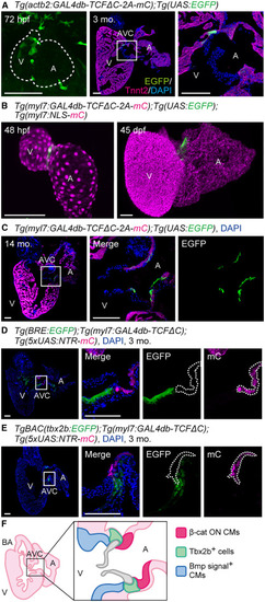
Myocardial Wnt/β-catenin signal-activated zone exists at the AVC in embryonic and adult stages (A) Representative confocal images of the hearts of Tg(actb2:GAL4db-TCFΔC-2A-mCherry);Tg(UAS:EGFP) fish. A ventral view of the heart at 72 h post-fertilization (hpf) (left). A cryosection image of the heart at 3 months stained with anti-GFP and anti-troponin T type 2 (Tnnt2) antibodies and 4′,6-diamidino-2-phenylindole (DAPI) (center). The boxed region is enlarged (right). Dotted lines outline the heart. Images shown in all figures are 3D-rendered confocal images of a stack unless otherwise noted. Confocal images in all figures are representative of at least three individuals. (B) Representative images of the hearts of Tg(myl7:GAL4db-TCFΔC-2A-mCherry);Tg(UAS:EGFP);Tg(myl7:NLS-mCherry) fish. A ventral view of the heart at 48 hpf (left). A resected heart at 45 days post-fertilization (dpf) was made transparent with clear, unobstructed brain/body imaging cocktails and computational analysis reagent-1A (CUBIC-1A) (right). Tg(myl7:NLS-mCherry) was used to outline the heart shape because the fluorescence of myl7:NLS-mCherry is brighter than that of myl7:GAL4db-TCFΔC-2A-mCherry. (C) A representative cryosection image of the heart of a 14-month-old Tg(myl7:GAL4db-TCFΔC-2A-mCherry);Tg(UAS:EGFP) fish stained with DAPI (left). The boxed region is enlarged (center and right). The fluorescence channel is indicated at the top. (D) A representative cryosection image of the heart of a 3-month-old Tg(BRE:EGFP);Tg(myl7:GAL4db-TCFΔC);Tg(5xUAS:NTR-mCherry) fish stained with DAPI (left). The boxed region is enlarged (right). Bmp signal+ cells do not overlap with β-cat ON CMs (dotted lines). (E) A representative cryosection image of the heart of a TgBAC(tbx2b:EGFP);Tg(myl7:GAL4db-TCFΔC);Tg(5xUAS:NTR-mCherry) fish at 3 months stained with DAPI (left). The boxed region is enlarged (right). Tbx2b+ cells do not overlap with β-cat ON CMs (dotted lines). (F) Schematic zonation at the AVC demarcated by the markers. β-cat ON CMs, Tbx2b+ cells, and Bmp signal+ cells were observed in the CMs at the atrial side of the AVC, at the root of the valves, and on the ventricular side of the AVC, respectively. Scale bars, 100 μm. A, atrium; AVC, atrioventricular canal; BA, bulbus arteriosus; CM, cardiomyocyte; mo, month; V, ventricle. See also Figure S1 and Videos S1A and S1B. |
Reprinted from Developmental Cell, 60(1), Chiba, A., Yamamoto, T., Fukui, H., Fukumoto, M., Shirai, M., Nakajima, H., Mochizuki, N., Zonated Wnt/β-catenin signal-activated cardiomyocytes at the atrioventricular canal promote coronary vessel formation in zebrafish, 21-29.e8, Copyright (2024) with permission from Elsevier. Full text @ Dev. Cell