
Fig. 2
- ID
- ZDB-FIG-250128-53
- Publication
- Gimenez et al., 2024 - Cohesin rad21 mutation dysregulates erythropoiesis and granulopoiesis output within the whole kidney marrow of adult zebrafish
- Other Figures
- All Figure Page
- Back to All Figure Page
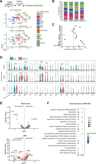
Single-cell RNA sequencing reveals the hematopoietic composition in WKM of adult wild type and rad21+/− zebrafish. A: uniform manifold projection (UMAP) showing the 22 cell clusters identified in scRNA-seq of WKM cells from wild type (WT) and rad21+/− adult zebrafish. B: stacked bar plot showing the percentage of annotated cell population within each genotype per replicate. WT1: 8,125 cells, WT2: 9,108 cells, rad21+/− 1: 6,159 cells, rad21+/− 2: 8,339 cells. C: fold change in population frequency in rad21+/− compared with WT. Red dot signifies statistically significant (P ≤ 0.05) change. Significance was determined using Student’s t test. D: violin plots of rad21, pcna, and myca expression in rad21+/− and WT. In rad21+/− versus WT comparison significance is from the scRNA-seq DESeq2 Wald test (Supplemental Table S3). *P < 0.05, ***P < 0.001, ****P < 0.0001. E: volcano plots showing DEGs identified in the comparison of rad21+/− versus WT in HSC-thromb and MPP clusters. Red dot signifies statistically significant (adjusted P ≤ 0.05) DEGs. F: GO enrichment analyses on up and down significant DEGs identified in MPP. Pathway analyses were performed using ClusterProfiler R package. Complete list of pathways and associated genes is provided in Supplemental Table S4. B, B-lymphocyte cells; E, erythroid; EP, erythroid progenitors; GMP, granulocytes monocyte progenitors; Gr, granulocytes; HSC, hematopoietic stem cell; M, monocytes/macrophages; MPP, multipotent progenitors; NK, natural killer; T, T-lymphocyte cells; thromb, thrombocytes; WKM, whole kidney marrow. |