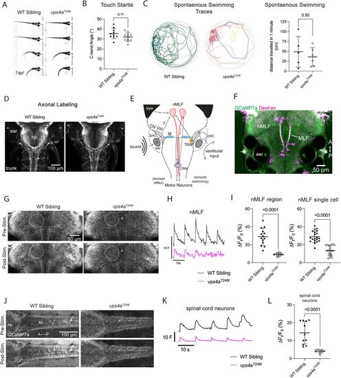
vps4aT248I mutants display defects in sensorimotor transformation. A, 7 dpf vps4aT248I mutant larvae have an intact startle reflex in response to aversive touch stimuli. B, 7 dpf vps4aT248I mutants (N = 11) do not have a significantly different C-bend angle compared with wild-type siblings (N = 10); unpaired t test; p = 0.1079; t = 1.687; df = 19. C, Traces of spontaneous swimming from 6 dpf wild-type siblings and mutants over the course of 1 min. vps4aT248I larvae (N = 5) swim shorter distances compared with wild-type siblings (N = 6); unpaired t test; p = 0.5063; t = 0.6921; df = 9) D, Acetylated tubulin antibody staining of vps4aT248I mutants shows no gross morphological defects in hindbrain/spinal cord or somatosensory axons. E, Simplified schematic showing the flow of information from a tone stimulus from the inner ear to motor neurons via Mauthner cell and nMLF pathways. S, saccular otolith; SHC, saccular hair cells; CN VIII, VIIIth nerve; M, Mauthner cell; CRN, cranial relay neurons; nMLF, nucleus of the medial longitudinal fasciculus; TAN, tangential neurons; UHC, utricular hair cells. F, 5 dpf larva showing basal GCaMP7a expression and backfill injected rhodamine dextran marking single cells within the nMLF. The nMLF region is outlined with a dotted line. G, Pre- and poststimulus GCaMP7a levels in the nMLF in a representative wild-type sibling and vps4aT248I mutant. H, Average trace of normalized fluorescence (F) for the nMLF of wild-type siblings and vps4aT248I mutants during tone stimulation at 600 Hz. I, vps4aT248I mutants (N = 11) have a significantly reduced percentage change fluorescence in the nMLF compared with wild-type siblings (N = 14); unpaired t test; p < 0.0001; t = 6.937; df = 23. Single backfilled neurons of the nMLF in vps4aT248I mutants (N = 18 cells from 6 larvae) also have a reduced change in percentage fluorescence compared with wild-type siblings (N = 20 cells from 9 larvae); unpaired t test; p < 0.0001; t = 8.018; df = 36. J, Pre- and poststimulus GCaMP7a levels in the spinal cord motor neurons of representative wild-type sibling and vps4aT248I mutant. K, Average trace of normalized fluorescence (F) for the spinal cord motor neurons of wild-type siblings and vps4aT248I mutants during tone stimulation at 450 Hz. L, vps4aT248I mutants (N = 9) have a significantly reduced percentage change fluorescence in spinal cord neurons compared with wild-type siblings (N = 11), Mann–Whitney test; p < 0.0001 (exact).
|
