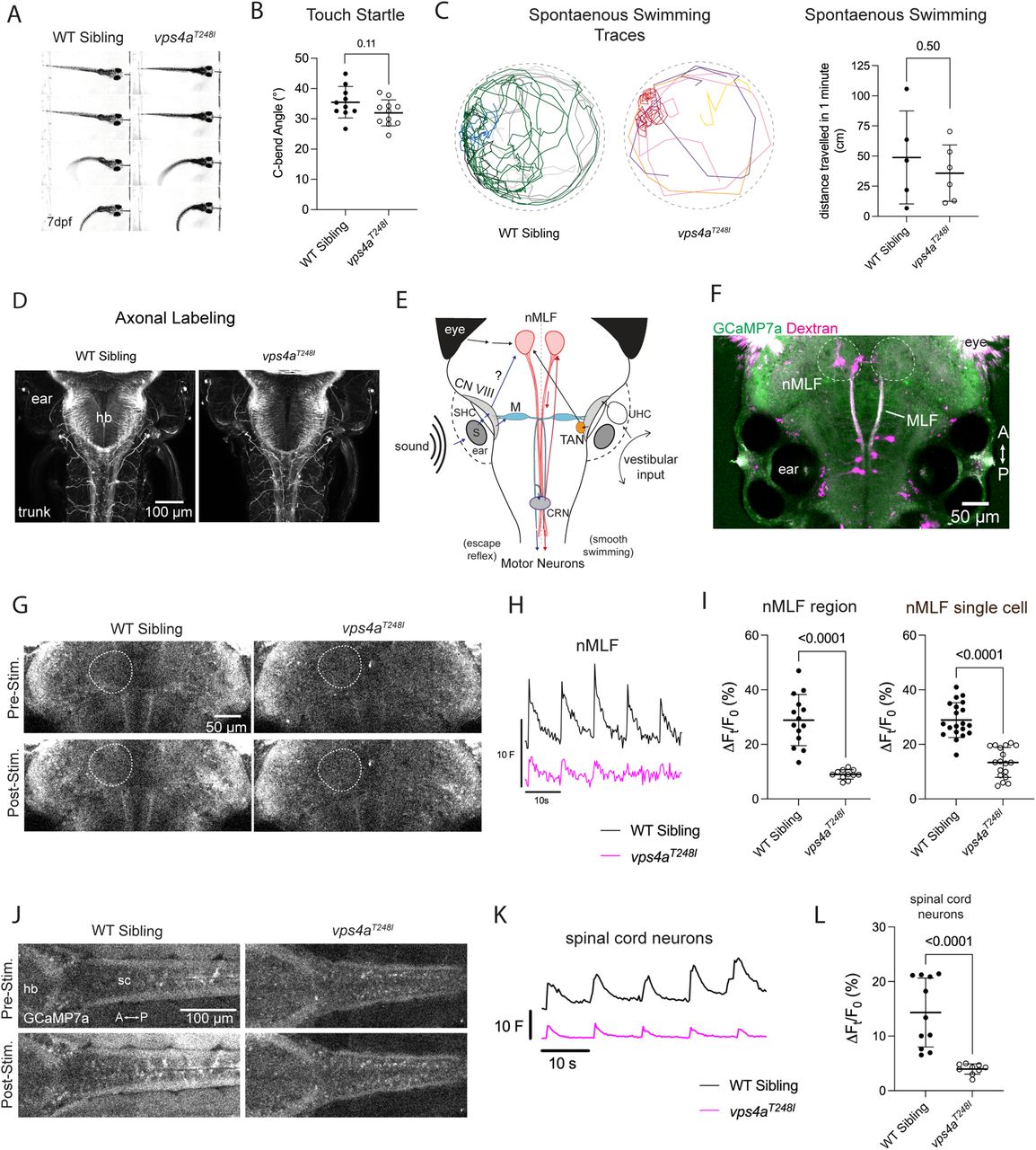
Fig. 7 vps4aT248I mutants display defects in sensorimotor transformation. A, 7 dpf vps4aT248I mutant larvae have an intact startle reflex in response to aversive touch stimuli. B, 7 dpf vps4aT248I mutants (N = 11) do not have a significantly different C-bend angle compared with wild-type siblings (N = 10); unpaired t test; p = 0.1079; t = 1.687; df = 19. C, Traces of spontaneous swimming from 6 dpf wild-type siblings and mutants over the course of 1 min. vps4aT248I larvae (N = 5) swim shorter distances compared with wild-type siblings (N = 6); unpaired t test; p = 0.5063; t = 0.6921; df = 9) D, Acetylated tubulin antibody staining of vps4aT248I mutants shows no gross morphological defects in hindbrain/spinal cord or somatosensory axons. E, Simplified schematic showing the flow of information from a tone stimulus from the inner ear to motor neurons via Mauthner cell and nMLF pathways. S, saccular otolith; SHC, saccular hair cells; CN VIII, VIIIth nerve; M, Mauthner cell; CRN, cranial relay neurons; nMLF, nucleus of the medial longitudinal fasciculus; TAN, tangential neurons; UHC, utricular hair cells. F, 5 dpf larva showing basal GCaMP7a expression and backfill injected rhodamine dextran marking single cells within the nMLF. The nMLF region is outlined with a dotted line. G, Pre- and poststimulus GCaMP7a levels in the nMLF in a representative wild-type sibling and vps4aT248I mutant. H, Average trace of normalized fluorescence (F) for the nMLF of wild-type siblings and vps4aT248I mutants during tone stimulation at 600 Hz. I, vps4aT248I mutants (N = 11) have a significantly reduced percentage change fluorescence in the nMLF compared with wild-type siblings (N = 14); unpaired t test; p < 0.0001; t = 6.937; df = 23. Single backfilled neurons of the nMLF in vps4aT248I mutants (N = 18 cells from 6 larvae) also have a reduced change in percentage fluorescence compared with wild-type siblings (N = 20 cells from 9 larvae); unpaired t test; p < 0.0001; t = 8.018; df = 36. J, Pre- and poststimulus GCaMP7a levels in the spinal cord motor neurons of representative wild-type sibling and vps4aT248I mutant. K, Average trace of normalized fluorescence (F) for the spinal cord motor neurons of wild-type siblings and vps4aT248I mutants during tone stimulation at 450 Hz. L, vps4aT248I mutants (N = 9) have a significantly reduced percentage change fluorescence in spinal cord neurons compared with wild-type siblings (N = 11), Mann–Whitney test; p < 0.0001 (exact).
Image
Figure Caption
Figure Data
Acknowledgments
This image is the copyrighted work of the attributed author or publisher, and
ZFIN has permission only to display this image to its users.
Additional permissions should be obtained from the applicable author or publisher of the image.
Full text @ J. Neurosci.

