
FIGURE
Fig. 8
Fig. 8
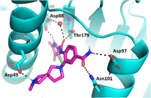
Docking binding mode of compound (R)-24e (in magenta sticks) in the ATP-binding site of Hsp90β (in cyan cartoon, PDB entry: 5UCJ). For clarity, only selected amino acid residues are presented as sticks. Hydrogen bonds are presented as black dashed lines. Structural water molecule is shown as a red sphere. |
Expression Data
Expression Detail
Antibody Labeling
Phenotype Data
Phenotype Detail
Acknowledgments
This image is the copyrighted work of the attributed author or publisher, and
ZFIN has permission only to display this image to its users.
Additional permissions should be obtained from the applicable author or publisher of the image.
Full text @ Eur. J. Med. Chem.