
Fig. 4
- ID
- ZDB-FIG-241105-30
- Publication
- Ceci et al., 2024 - RACK1 contributes to the upregulation of embryonic genes in a model of cardiac hypertrophy
- Other Figures
- All Figure Page
- Back to All Figure Page
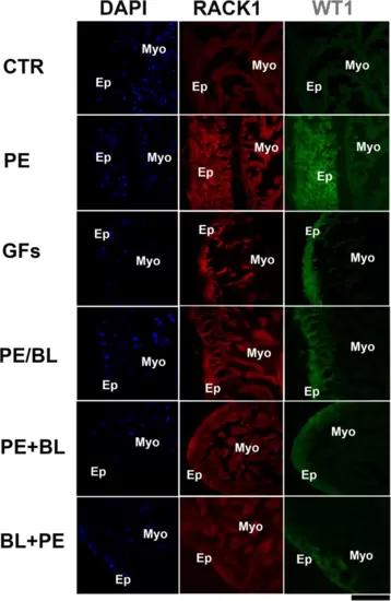
Double staining of RACK1 and WT1 was performed via confocal microscopy. RACK1 (red fluorescence) is expressed at lower levels in the epicardium than in the myocardium in the CTR. In all the experimental groups, RACK1 expression was increased, and RACK1 was particularly colocalized with the WT1 antibody, which stains the epicardium (green fluorescence). WT1 is strongly expressed in PE and GFs and moderately expressed in PE + BL, PE/BL, and BL + PE. The hearts utilized for the experiments were N = 4/5 sections in each group in triplicate experiments. Blue fluorescence: DAPI (nuclear marker); scale bar: 500 μm. |