FIGURE

Figure 8

ID
ZDB-FIG-241019-69
Publication
Lee et al., 2024 - The 419th Aspartic Acid of Neural Membrane Protein Enolase 2 Is a Key Residue Involved in the Axonal Growth of Motor Neurons Mediated by Interaction between Enolase 2 Receptor and Extracellular Pgk1 Ligand
Other Figures
All Figure Page
Back to All Figure Page
Figure 8

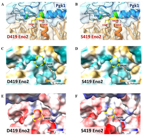
Molecular docking model to illustrate the key amino acid involved in Eno2-ePgk1 interaction. (A,B) Simulated model to illustrate how Eno2 (PDB ID: 5TD9) interacted with ePgk1 (PDB ID: 2ZGV) is presented in blue and orange, respectively. Drawing of partial enlargement of critical amino acid residues involved in Eno2-ePgk1 interaction was presented, including (A) wild-type Eno2 D419 and (B) mutant Eno2 D419S. The segment of the 345th to 360th amino acids of Pgk1, the segment of the 404th to 431st amino acid of Eno2, and the mutant Eno2 D419th were shown in Dodger blue, chocolate, and orange, respectively. (C,D) Drawing of partial enlargement of the surface polarity distribution of (C) Eno2 D419 and (D) Eno2 D419S. Dotted circles highlighted the D419th residue on Eno2. Lipophilicity was presented as low (hydrophilicity, in cyan) to high (hydrophobicity, in brown). Dotted zone indicates the 419th site. (E,F) Drawing of partial enlargement of the surface charge distribution of (E) Eno2 D419 and (F) Eno2 D419S. Chargeability from negative charge (in red) to positive charge (in blue). Dotted circles marked the 419th residue of Eno2.

Expression Data

Expression Detail
Antibody Labeling
Phenotype Data

Phenotype Detail
Acknowledgments
This image is the copyrighted work of the attributed author or publisher, and ZFIN has permission only to display this image to its users. Additional permissions should be obtained from the applicable author or publisher of the image. Full text @ Int. J. Mol. Sci.