|
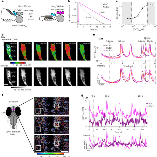
Quantitative Ca2+ measurements by FLIM using WHaloCaMP1a. a, Schematic of WHaloCaMP1a bound to a dye-ligand used as a FLIM probe. Tryptophan quenching modulates the fluorescence lifetime. b, Normalized fluorescence lifetime of WHaloCaMP1a669 in the presence or the absence of Ca2+, fit to a three-component fluorescence decay. c, Calibration curve of the averaged fluorescence lifetime of WHaloCaMP1a669 versus Ca2+ concentration. The white box indicates the range in which WHaloCaMP1a669 can be used to make quantitative measurements of Ca2+ concentration. Performed with purified protein. Mean of three replicates and s.d. are plotted. d, Pseudocolored concentration (top) and intensity images (bottom) of WHaloCaMP1a669 in HeLa cells after histamine addition. Scale bar, 20 µm. Color bar indicates Ca2+ concentration, calculated from a calibration curve of fluorescence lifetime. AU, arbitrary units. e, Quantitative Ca2+ concentration calculated (calc) from a FLIM calibration curve (top) and fluorescence traces ∆F/F0 calculated from the intensity channel (bottom) in histamine-stimulated HeLa cells in the ROI highlighted in d. Calibrated WHaloCaMP1a669 can only be used to measure Ca2+ concentrations up to 200 nM, indicated by a dashed horizontal line. Vertical dashed lines indicate time points in the time series at which images in d are shown. f, FLIM of WHaloCaMP1a669 in live zebrafish larvae showing spontaneous neuronal activity in the forebrain. The experiment was repeated independently three times with similar results. Schematic indicating the field of view during imaging (left). Overlaid images of FLIM and intensity using Leica LAS X software, with a color bar indicating the fluorescence lifetime. Scale bar, 20 µm. g, Ca2+ concentrations calculated from a FLIM calibration curve (top) and fluorescence traces ∆F/F0 calculated from the intensity channel (bottom) over time for two neurons in the forebrain of zebrafish larvae from the ROI indicated in f. Dashed lines indicate time points of images in f. Representative images from three imaging sessions.
|