|
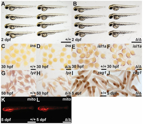
Investigation of the socs4bΔ18 mutants. Light microscopy of homozygous socs4b WT (+/+; (A)) and socs4bΔ18 (Δ/Δ; (B)) embryos at 2 dpf. Homozygous socs4b WT (+/+; (C,E,G,I,K)) and socs4bΔ18 (Δ/Δ; (D,F,H,J,L)) embryos subjected to WISH at the indicated times with specific probes for ins ((C), n = 35; (D), n = 33), isl1a ((E), n = 42; (F), n = 38), lyz ((G), n = 27; (H), n = 30), and rag1 ((I), n = 40; (J), n = 37) viewed by light microscopy or staining with Mitotracker Red (mito) ((K), n = 23; (L), n = 25) and viewed by fluorescence microscopy. The number of embryos analyzed with each stain is shown. Scale bars = 1 mm.
|