
Figure 3
- ID
- ZDB-IMAGE-240930-148
- Genes
- Publication
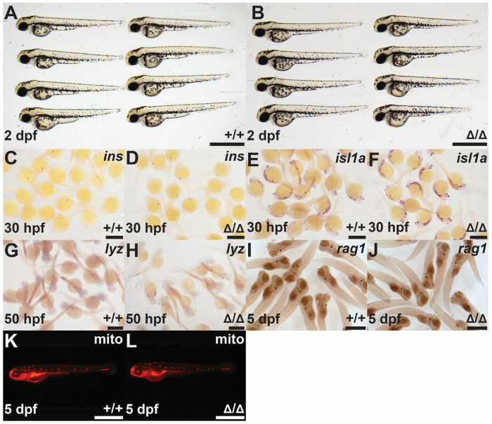
- Trengove et al., 2024 - Zebrafish Suppressor of Cytokine Signaling 4b (Socs4b) Is Dispensable for Development but May Regulate Epidermal Growth Factor Receptor Signaling
- All Figures
- Figures for Trengove et al., 2024
|
Figure 3
Investigation of the