
Fig. 8
- ID
- ZDB-FIG-240912-78
- Publication
- Boldizar et al., 2024 - The role of cilia in the development, survival, and regeneration of hair cells
- Other Figures
- All Figure Page
- Back to All Figure Page
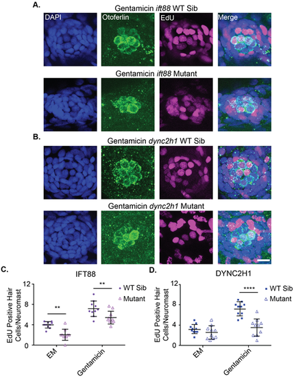
Proliferation during hair cell regeneration is reduced in IFT gene mutants. Representative images of neuromasts from (A) ift88 and (B) dync2h1 wild-type siblings (top) and mutants (bottom) at 7 dpf following gentamicin and EdU treatment from 4-5 dpf. Cells that proliferated during the 4-5 dpf time period are EdU-positive as shown in magenta. Nuclei are labeled in blue with DAPI and hair cells in green with the otoferlin antibody. Scale bar: 10 µm. The graphs show the quantification of EdU-positive hair cells/neuromast for (C) ift88 and (D) dync2h1 wild-type siblings and mutants 48 h after either EM or gentamicin treatment. A two-way ANOVA was carried out for both experiments. For ift88 both drug treatment and genotype were significant sources of variation (P<0.0001) whereas the interaction between the two variables was not (P=0.7884). For dync2h1 both of the individual variables were also significant sources of variation (P<0.0001) as was the interaction between the two variables (P=0.0016). **=P<0.01 and ****=P<0.0001 by Tukey's multiple comparisons test comparing the two groups at the edges of the lines under the asterisks. n=10 for all groups. |