
Fig. 2
- ID
- ZDB-FIG-240903-172
- Publication
- Casey et al., 2024 - A simple and scalable zebrafish model of Sonic hedgehog medulloblastoma
- Other Figures
- All Figure Page
- Back to All Figure Page
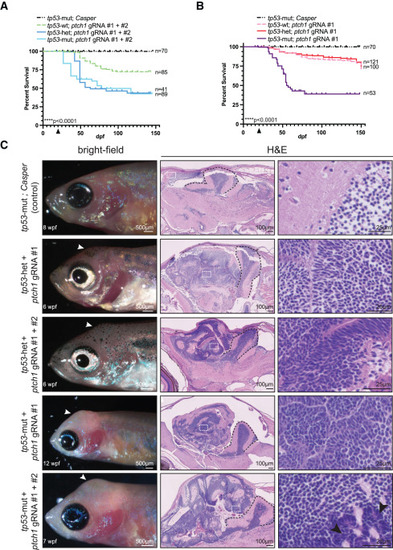
Mutation in tp53 promotes aggressiveness in ptch1-crispant brain tumors (A and B) Zebrafish from the indicated genetic backgrounds were injected at the one-cell stage with either ptch1 gRNAs #1 and #2 (A) or ptch1 gRNA #1 alone (B) and analyzed for survival following injection. The arrowhead on the x axis indicates the beginning of survival analysis. The total number of animals per group is indicated on the right. Data are plotted from a single experiment except for tp53-mut conditions, where data had to be pooled from multiple experiments to generate enough animals. Experiments were repeated at least twice. Statistical significance was calculated using the Mantel-Cox test. ∗∗∗∗p < 0.0001. (C) (Left) Bright-field images of 6–12 wpf animals with the indicated genetic backgrounds that were injected at the one-cell stage with either ptch1 gRNA #1 alone or ptch1 gRNAs #1 and #2. (Middle and right) Sagittal sections of control brain or ptch1-crispant brain tumors stained with H&E. White arrowheads indicate location of the tumor in the left. Black arrowheads in the lower right indicate larger neoplastic cells with more nuclear molding. White boxes in the middle are shown at higher magnification in the right. Black dashed lines in the middle indicate the cerebellum. See also Figure S1 and Table S1. Adult germline ptch1 mutants develop brain tumors To independently verify that loss of ptch1 causes brain tumors in zebrafish, we next tested whether germline loss of ptch1 also gives rise to brain tumors. Due to the short lifespan and compromised health of the transient ptch1-crispant adult animals (Figure 1), it was not possible to establish a stable line using our ptch1-crispant methodology. We therefore obtained and analyzed a previously established ptch1tj222 line that has a premature stop codon at position 590 (Figures 1A and 3) and has primarily been studied during embryonic development.38,39 As expected from previous reports, most of the ptch1tj222 homozygotes died at early developmental stages; however, we identified four homozygous mutant escapers out of 147 adults (∼37 homozygotes would be expected with normal Mendelian ratios) from an incross between ptch1tj222 heterozygotes. All four homozygous mutants developed brain tumors that resemble ptch1-crispant tumors at the morphological level, and the ability to form tumors was independent of tp53 status (Figure 3A). Analysis of the germline ptch1-null allele supports the conclusion that loss of ptch1 drives the development of brain tumors in zebrafish. |