
Fig. 5
- ID
- ZDB-FIG-240828-48
- Publication
- De Bortoli et al., 2024 - Somatic loss-of-function PIK3R1 and activating non-hotspot PIK3CA mutations associated with Capillary Malformation with Dilated Veins (CMDV)
- Other Figures
- All Figure Page
- Back to All Figure Page
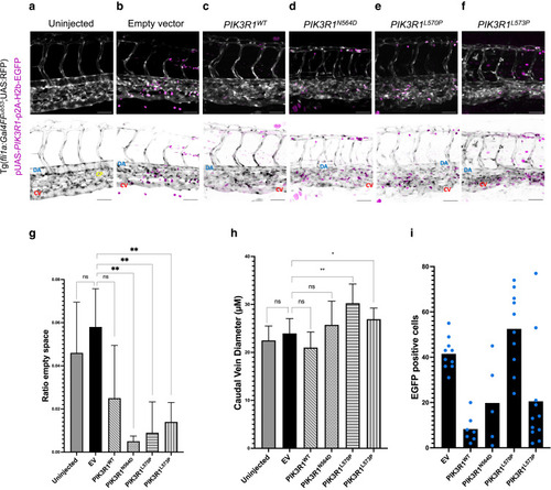
Endothelial-specific overexpression of mutated PIK3R1 in Tg(fli1a:Gal4FFub53;UAS:RFP)-positive zebrafish embryos. Normal and inverted images are shown. EGFP+ cells (magenta) contain the injected plasmid. CV (red), DA (yellow), and CP (blue) are shown. (a) Embryos with normal development of trunk vasculature. (b, c) Embryos injected with pUAS-p2A-H2b-EGFP (EV) and pUAS-PIK3R1WT-p2A-H2b-EGFP: normal development of trunk vasculature. (d–f) Embryos injected with pUAS-PIK3R1-p2A-H2b-EGFP expressing mutated PIK3R1: CV enlargement and CP disruption. (g) Quantification of area between CV and DA occupied by the capillary bed: ratio between intravessel and intervessel space. (h) Quantification of CV diameter. (i) Quantification of plasmid integration rate in zebrafish endothelium: EGFP+ cells found in capillary–venous plexus. Plotted are averages normalized on the number of embryos for each condition. Each blue dot corresponds to 1 embryo. Bar = 50 μM. Error bars are shown as measure of variability and represent the SD. Number of biological replicates: uninjected, n = 19; EV, n = 10; PIK3R1WT, n = 9; PIK3R1N564D, n = 4; PIK3R1L570P, n = 8; and PIK3R1L573P, n = 9. Embryos in which the CV was not readily distinguishable were discarded. CV, caudal vein; CP, capillary plexus; DA, dorsal aorta; EV, empty vector; ns, not significant. |