
Fig. 4
- ID
- ZDB-FIG-240828-47
- Publication
- De Bortoli et al., 2024 - Somatic loss-of-function PIK3R1 and activating non-hotspot PIK3CA mutations associated with Capillary Malformation with Dilated Veins (CMDV)
- Other Figures
- All Figure Page
- Back to All Figure Page
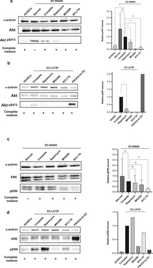
Western blot analysis of Akt and ERK phosphorylation and treatment with PI3K pathways inhibitors. (a) Patient-derived EC-N564D display Akt phosphorylation. pAkt is decreased after treatment with rapamycin and BYL719 (48 h) or MK2206 (24 h). (b) Patient-derived EC-L573P display Akt phosphorylation. pAkt is decreased after treatment with rapamycin and BYL719 (48 h) or MK2206 (24 h). (c) Patient-derived EC-N564D show ERK phosphorylation. pERK is decreased upon treatment with BYL719 but not with rapamycin or MK2206. (d) Patient-derived EC-L573P show ERK phosphorylation. pERK is decreased upon treatment with BYL719 and MK2206 but not with rapamycin. α-Actinin was used as loading control. PIK3CA-hs ECs are primary cells from a patient with CLVM (H1047R PIK3CA mutation). Error bars are shown as measure of variability and represent the SD. Experiments were repeated n = 5 times for EC-N564D cells. Owing to a shortage of surgical resected tissue and limited number of viable EC-L573P passages, Akt/pAkt and ERK/pERK were repeated n = 2 and n = 1 times, respectively. Akt, protein kinase B; CLVM, capillaro–lymphatico–venous malformation; EC, endothelial cell; ERK, extracellular signal–regulated kinase; h, hour; hs, hotspot; ns, not significant; pAkt, phosphorylated protein kinase B; pERK, phosphorylated extracellular signal–regulated kinase; PI3K, phosphoinositide 3-kinase. |