
Fig. 7
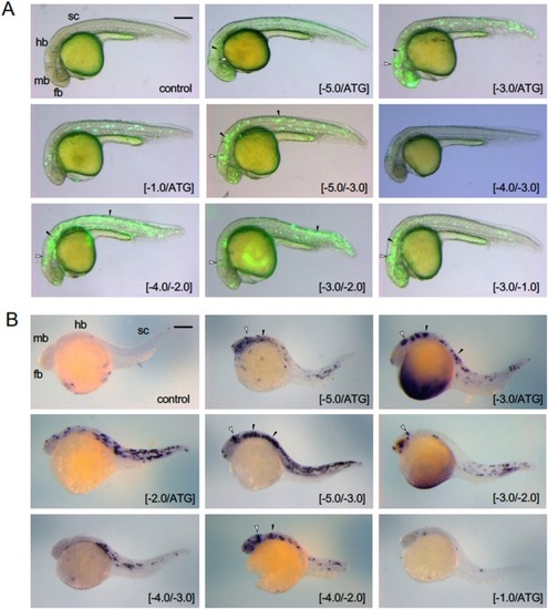
Expression of egfp following co-injection with pax2a upstream DNA. DNA sub-fragments from the upstream DNA of pax2a ( Fig. 6 A) were co-injected with hsp-egfp DNA into fertilized eggs, which were examined at approximately 24 hpf for (A) EGFP fluorescence ( Fig. 6 B) and (B) egfp mRNA visualized by WISH. Co-injected sub-fragments are shown at the bottom right. Reporter expression in the MHB and the ventral hindbrain/spinal cord is marked with open triangles and black triangles, respectively. Regarding WISH, all sub-fragments were examined in at least 10 embryos. Due to the mosaicism of transient transgene expression, the patterns were diverse, but typical data are selected and shown. Additional data showing lack of reporter expression are provided in Fig. S5 . fb, forebrain; hb, hindbrain; mb, midbrain; sc, spinal cord. Scale bar, 250 μm. |