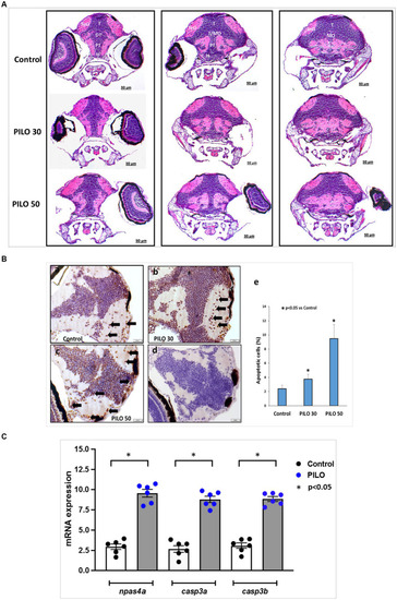
Histological changes in the brain of zebrafish larvae exposed to pilocarpine (PILO). (A) Brain morphology under hematoxylin and eosin (H&E) staining—larvae were incubated in medium containing PILO (30 or 50 mM) for 2 h. Next, larvae were thoroughly washed out × 5 times and incubated in fresh medium for subsequent 22 h. Afterwards, larvae were cooled down and fixed in 10% buffered formalin for 24 h at room temperature and processed for further analysis. DT, dorsal thalamus; H, hypothalamus; MO, medulla oblongata; PT, posterior tuberculum; T, midbrain tegmentum; TeO, tectum opticum; T/MO, midbrain tegmentum/medulla oblongata boundary. (B) Apoptosis evaluated by means of TUNEL staining. Larvae were incubated in medium containing PILO (30 or 50 mM) for 2 h. Next, larvae were thoroughly washed out × 5 times and incubated in fresh medium for subsequent 22 h. Afterwards, larvae were cooled down and fixed in 10% buffered formalin for 24 h at room temperature and processed for further analysis: (a–c) TUNEL staining, arrows indicate representative TUNEL-positive cells marked as dark brown; (d) a negative control was performed without active TdTenzyme; (e) the percentage of apoptotic cells over the total cell number per section per larva—the data are presented as the means ± standard deviations (SDs) (N = 5/group)—a one-way ANOVA test, followed by Dunnett’s multiple comparison analysis, were used for statistical evaluation (the significance was set at p < 0.05). (C) mRNA expression of npsa4a (marker of neuronal activity), casp3a and casp3b (markers of apoptosis). Larvae were incubated in medium containing PILO (50 mM) for 2 h. Next, larvae were thoroughly washed out × 5 times and incubated in fresh medium for subsequent 22 h. Next, samples were collected (n = 6 per group, N = 25 larvae per sample).
|
