|
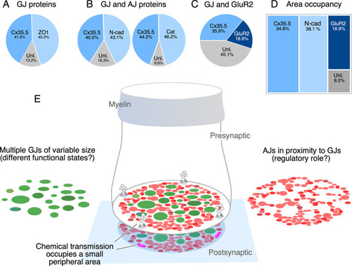
The electrical synapse at the club ending (CE) combines multiple gap junctions (GJs) with adherens junctions (AJs). (A) Double labeling with anti-Cx35.5 and anti-ZO1 shows a similar proportion of fluorescence at CEs (ZO1 45.24% ± 2.709; Cx35.5 41.53% ± 4.058; unlabeled 13.23%; n = 8 CEs from six fish). The region of interest (ROI) for analysis of fluorescence here and (B) was defined by the outline of Cx35.5 labeling of CEs. (B) Double labeling for N-cadherin and Cx35.5 (left), and for β-catenin and Cx35 (right) also shows similar proportionality (N-Cad 43.20% ± 3.334; Cx35.5 40.48% ± 3.041; unlabeled 16.32%; n = 13 from five fish; β-catenin 46.21% ± 2.728; Cx35.5 44.18% ± 1.671; unlabeled 9.61%; n = 13 CEs from seven fish). (C) Double labeling for Cx35.5 and GluR2 shows lack of proportionality, with Cx35.5 fluorescence occupying the majority of the CE contact area (GluR2 18.99% ± 1.601; Cx35.5 35.92% ± 2.087; unlabeled 45.10%; n = 10 CEs from five fish). ROI was defined in this case by the outline of GluR2 labeling. (D) Tree plot illustrating the area occupancy (fluorescence/contact area) for AJ (N-Cad = 38.1%), GJ (Cx35 = 34.8%), and glutamatergic (GluR2=18.9%) labeling at individual CEs (values normalized to ROI outlined by GluR2 labeling). The unlabeled area represents 8.1% of the contact’s surface. (E) The cartoon summarizes the synaptic components identified at a single CE contact. While chemical synapses are restricted to a small and peripheral area of the contact (presynaptic vesicles and release sites are represented in gray, postsynaptic receptor areas in magenta), most of its contact surface is occupied by multiple GJs (green) of variable size, which are interleaved and closely associated to AJs (red).
|