
Figure 1.
- ID
- ZDB-FIG-240820-1
- Publication
- Cárdenas-García et al., 2024 - The components of an electrical synapse as revealed by expansion microscopy of a single synaptic contact
- Other Figures
- All Figure Page
- Back to All Figure Page
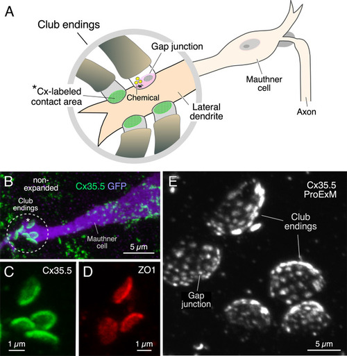
Expansion microscopy of club ending (CE) contact areas in larval zebrafish. (A) The cartoon illustrates the auditory afferents that terminate as single CEs, each containing gap junctions (GJs, green) and specializations for chemical transmission (Chemical), on the distal portion of the lateral dendrite of the Mauthner (M-) cell. Synaptic contact areas labeled with connexin antibody (see B and C) are represented in green. (B) Confocal image with anti-GFP (purple) and anti-Cx35/36 (green), which labels both Cx35.5 and Cx35.1, showing a long stretch of the lateral dendrite of the M-cell (projection of 34 confocal z-sections at 0.39 µm z-step size), revealing the contact areas (indicated by the asterisk here and the cartoon of A) of several CEs. (C, D) Contact areas of individual CEs labeled with anti-Cx35/36 (C, green; projection of 12 sections at 0.39 µm z-step size) and anti-ZO1 (D, red; projection of 4 sections at 0.39 µm z-step size). (E) Protein-retention expansion microscopy (ProExM) with anti-Cx35.5 increases the size of CE synaptic contact areas, enabling the visualization of intrasynaptic components (projection of 19 sections at 0.88 µm z-step size). The scale bars represent actual dimensions and, therefore, ProExM images were not adjusted for expansion factor. |