
FIGURE
Fig. 2
- ID
- ZDB-FIG-240720-2
- Publication
- de Sousa et al., 2024 - Efficacy of the bumped kinase inhibitor BKI-1708 against the cyst-forming apicomplexan parasites Toxoplasma gondii and Neospora caninum in vitro and in experimentally infected mice
- Other Figures
- All Figure Page
- Back to All Figure Page
Fig. 2
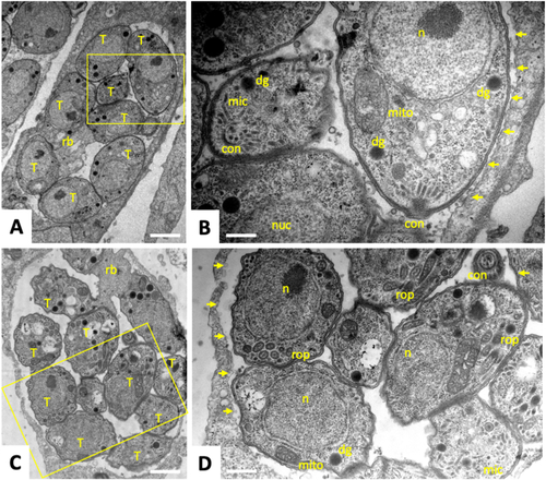
Transmission Electron Microscopy of |
Expression Data
Expression Detail
Antibody Labeling
Phenotype Data
Phenotype Detail
Acknowledgments
This image is the copyrighted work of the attributed author or publisher, and
ZFIN has permission only to display this image to its users.
Additional permissions should be obtained from the applicable author or publisher of the image.
Full text @ Int J Parasitol Drugs Drug Resist