
FIGURE
Fig. 5
- ID
- ZDB-FIG-240715-90
- Publication
- Ligunas et al., 2024 - Tissue-specific and endogenous protein labeling with split fluorescent proteins
- Other Figures
- All Figure Page
- Back to All Figure Page
Fig. 5
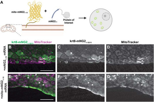
Directing protein localization with split-mNG2. A. Schematic illustrating use of the split-mNG2 system to sequester proteins of interest on mitochondria. B–G. Representative images of krt8-mNG211 embryos injected with mNG21-10 (B–D) or mito-mNG21-10 (E–G) mRNA and stained with MitoTracker dye to label mitochondria. Maximum projections of confocal z-stacks. Arrows indicate colocalization between split-mNG2 and MitoTracker fluorescence. Images were acquired from the tail fin epidermis at 48 h post-fertilization (hpf). Scale bars, 50 μm. |
Expression Data
Expression Detail
Antibody Labeling
Phenotype Data
Phenotype Detail
Acknowledgments
This image is the copyrighted work of the attributed author or publisher, and
ZFIN has permission only to display this image to its users.
Additional permissions should be obtained from the applicable author or publisher of the image.
Reprinted from Developmental Biology, 514, Ligunas, G.D., Paniagua, G., LaBelle, J., Ramos-Martinez, A., Shen, K., Gerlt, E.H., Aguilar, K., Nguyen, N., Materna, S.C., Woo, S., Tissue-specific and endogenous protein labeling with split fluorescent proteins, 109-116, Copyright (2024) with permission from Elsevier. Full text @ Dev. Biol.