
FIGURE
Fig. 5
- ID
- ZDB-FIG-240708-34
- Publication
- Warns et al., 2024 - scMultiome analysis identifies a single caudal hindbrain compartment in the developing zebrafish nervous system
- Other Figures
- All Figure Page
- Back to All Figure Page
Fig. 5
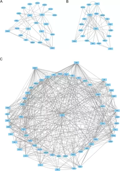
Caudal hindbrain cells display a distinct gene regulatory network at 13hpf. (A-C.) Direct-Net derived gene regulatory networks for r6 (A), CHB (B) and SC (C) at 13hpf. TFs (squares) and non-TFs (ovals) are linked by the presence of accessible TF motifs within 500 bp (solid lines) or 250 kb (dashed lines) of a gene’s transcription start site. Due to the large network produced for SC, only nodes with more than ten connections are shown (the full SC network is shown in Additional file 7, Figure S2B) |
Expression Data
Expression Detail
Antibody Labeling
Phenotype Data
Phenotype Detail
Acknowledgments
This image is the copyrighted work of the attributed author or publisher, and
ZFIN has permission only to display this image to its users.
Additional permissions should be obtained from the applicable author or publisher of the image.
Full text @ Neural Dev.