
Fig. 2
- ID
- ZDB-FIG-240708-31
- Publication
- Warns et al., 2024 - scMultiome analysis identifies a single caudal hindbrain compartment in the developing zebrafish nervous system
- Other Figures
- All Figure Page
- Back to All Figure Page
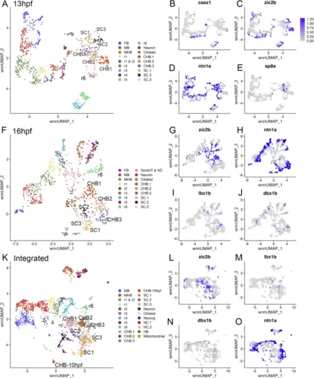
The caudal hindbrain can be subdivided along its dorsoventral axis. See also Additional file 2, Table S2. (A) UMAP of 13hpf neural clusters. (B-E) Feature plots showing expressing of the indicated genes. (F) UMAP of 16hpf neural clusters. (G-J) Feature plots showing expressing of the indicated genes. (K) UMAP of integrated 10hpf, 13hpf and 16hpf neural clusters. (L-O) Feature plots showing expressing of the indicated genes. UMAPs in A, F, K are based on 8.0, 6.0 and 5.0 res clustering, respectively (chosen because this fully resolves r1-r6). In cases where multiple clusters were assigned the same identity, they were combined into a single cluster with that identity – except for CHB and SC clusters that were left separate and labeled consecutively. See legend to Fig. 1 for abbreviations |