
Fig. 3
- ID
- ZDB-FIG-240620-63
- Publication
- Bongiovanni et al., 2024 - BMP7 promotes cardiomyocyte regeneration in zebrafish and adult mice
- Other Figures
- All Figure Page
- Back to All Figure Page
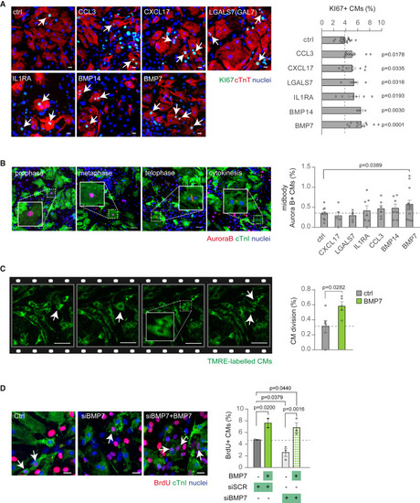
BMP7 robustly induces cell cycle activity and cell division in neonatal mammalian cardiomyocytes (A and B) Cardiomyocytes isolated from postnatal day 1 (P1) mice were cultured in vitro and stimulated for 48 h with selected growth factors (10 ng/mL), namely CCL3, CXCL17, GAL7 (LSGAL7), IL1RA, BMP14, and BMP7. Cardiomyocytes were identified by cardiac Troponin T (cTnT) or Troponin I (cTnI) staining and analyzed by immunofluorescence for (A) cell-cycle activity (KI67) or (B) cytokinesis (midbody Aurora B kinase) (n = 13,961 cardiomyocytes pooled from the analysis of 68 samples in A; n = 57,498 cardiomyocytes pooled from the analysis of 55 samples in B); representative pictures are provided; arrows point at proliferating cardiomyocytes; scale bars, 20 μm in (A) and 50 μm in (B). (C) Quantification and representative images of cell division events (n = 11 samples with a total of 5184 cardiomyocytes analyzed) in TMRE-labeled neonatal cardiomyocytes detected in 16-h time-lapse imaging at 15-min intervals in vitro; arrows point at cardiomyocytes undergoing cell division; scale bars, 50 μm. (D) Evaluation of neonatal (postnatal day 1, P1) cardiomyocyte proliferation by immunofluorescence analysis of DNA synthesis (BrdU assay) following knockdown of BMP7 and stimulation with/without BMP7 (10 ng/mL) for 48 h (n = 2,480 cardiomyocytes pooled from the analysis of 11 samples). Representative pictures are provided; scale bars, 20 μm; arrows point at cycling cardiomyocytes. The values are presented as mean (error bars show SEM), statistical significance was determined using one-way ANOVA followed by Sidak’s test in (A), (B), and (D) (comparison between pairs of treatments; control vs. selected growth factor) and two-sided Student’s t test in (C). |