|
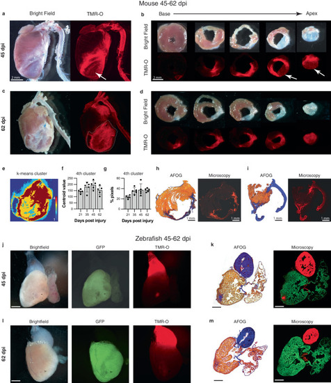
Dynamic deposition and oxidation of new collagen in chronic infarcts. a, b Whole-heart and short axis section of a mouse heart 45 dpi. Some areas of the remodeled scar are structurally static with little TMR-O uptake (arrows, n = 5). However, other areas of the scar show substantial amounts of TMR-O uptake, consistent with the oxidation of newly deposited collagen. c, d Similarly, at day 62 some areas of the scar show high amounts of probe uptake, consistent with dynamic collagen deposition (n = 5). e–g K-means clustering (4 clusters) of the infarct zone in the whole-heart TMR-O images. e TMR-O clusters in a day 62 infarct zone, demarcated by the dashed lines in panel C. The cluster of highest TMR-O signal (4th cluster, brown) occupies a substantial portion of the infarct. f, g The centroid value and area of the 4th cluster did not change significantly over time (n = 5, values are plotted as the average ± the standard deviation) one-way ANOVA, with Tukey’s multiple comparisons, consistent with active/new collagen deposition in the infarcts at all stages of healing. Source data are provided in the Source Data file. h, i AFOG staining, and fluorescence microscopy confirmed the heterogeneity of TMR-O uptake in the day 45 and 62 murine infarcts (n = 5). j, k In injured zebrafish hearts (n = 5, scale bar = 300 μm.) most of the myocardium had been regenerated by 45 dpi and the size of the residual injury was small. However, within these small residual areas of injury substantial levels of TMR-O uptake were still seen. l, m Within 2 months of injury no evidence of myocardial injury or TMR-O uptake was seen in the zebrafish hearts (n = 5, scale bar = 300 μm.), consistent with complete resorption of the scars and successful regeneration of the myocardium. Scale bars in (g–j) are 300 µm.
|