Fig. 5
- ID
- ZDB-FIG-240611-32
- Publication
- Ding et al., 2024 - Mycobacterial CpsA activates type I IFN signaling in macrophages via cGAS-mediated pathway
- Other Figures
- All Figure Page
- Back to All Figure Page
|
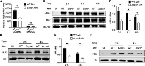
CpsA-related type I IFN production is through cGAS-TBK1-IRF3 pathway (A) BMDMs extracted from WT mice or Cgas knockout mice were infected with WT or ΔcpsA Mm strains at MOI of 10. RNA was isolated at 10 h after infection. Ifnβ levels were determined by RT-qPCR in different infection cells; values were normalized to Gapdh and uninfected BMDMs. (B) RAW264.7 cells were infected with WT and ΔcpsA strains at MOI of 10. Total cell lysates and nuclear part were harvested at indicated time points. Phospho-TBK1 and TBK1 protein levels were determined by using a densitometer and (C) corresponding statistical results were calculated from two independent experiments by ImageJ software, GAPDH as a reference protein. (D) The protein levels of IRF3 from nuclear fraction were determined by western blot (WB) and (E) corresponding statistical results were calculated from two independent experiments by ImageJ software, H3 as a reference. (F) Nuclear p65 and H3 protein levels were determined by WB. Shown is a representative experiment of three. Data represent means ± SD of three independent experiments. A, ∗p < 0.05, ns, not significant by unpaired t test, and C, D ∗p < 0.05, ∗∗p < 0.01, ns, not significant by two-way ANOVA with multiple comparisons test. |

