FIGURE

Fig. 5

ID
ZDB-FIG-240601-5
Publication
Xiao et al., 2024 - mTOR mutation disrupts larval zebrafish tail fin regeneration via regulating proliferation of blastema cells and mitochondrial functions
Other Figures
All Figure Page
Back to All Figure Page
Fig. 5

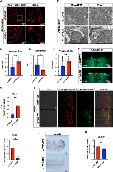
mTOR knock out affected mitochondrial morphology and membrane potential. (A) Mito-Tracker red staining of mTOR-WT and mTOR-KO larval zebrafish fin following amputation. (B) Representative transmission electron microscope images of mTOR-WT and mTOR-KO larval zebrafish fin after amputation. (C-E) Statistical analysis of the average area, aspect ratio and average width of mitochondria between mTOR-WT and mTOR-KO larval zebrafish fin after amputation. (F-G) The ROS levels between mTOR-WT and mTOR-KO larval zebrafish fin after amputation. (H-I) Measurement of the mitochondrial membrane potential by JC-1 staining between mTOR-WT and mTOR-KO zebrafish at 48 hpa. (J) Location of mitochondrial fission related gene (dnm1l) in mTOR-WT and mTOR-KO larval zebrafish fin by in situ hybridization. (K) mRNA expression level of dnm1l in mTOR-WT and mTOR-KO larval zebrafish fin. *P < 0.05, **P < 0.01

Expression Data

Expression Detail
Antibody Labeling
Phenotype Data

Phenotype Detail
Acknowledgments
This image is the copyrighted work of the attributed author or publisher, and ZFIN has permission only to display this image to its users. Additional permissions should be obtained from the applicable author or publisher of the image. Full text @ J Orthop Surg Res