
Fig. 2
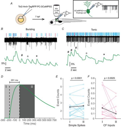
BOTH SIMPLE SPIKES AND CF EPSPS CONTRIBUTE TO THE CALCIUM SIGNAL IN PNS, IRRESPECTIVE OF CELLULAR STATE A, schematic of the experimental design and setup of simultaneous calcium imaging and electrophysiology in larval zebrafish. B and C, representative traces obtained during simultaneous imaging and electrophysiology for cells in the bursting (B) and tonic (C) mode. Simple spikes and CF inputs in the trace are identified and marked in the raster above the trace in cyan and magenta, respectively. The simultaneously-recorded change in GCaMP fluorescence (ΔF/F) is shown below in green. Calcium transients corresponding to simple spikes only (‘∗’), CF-events only (‘#’), and both events (‘o’) are marked. D, the typical GCaMP5G response to a single action potential, assuming it follows the profile of a difference of single exponentials with a half-rise time of 100 ms and half-decay time of 500 ms. The vertical dashed lines indicate the time of initiation of the calcium transient and the time when the transient peaks, which corresponds to an interval of 201 ms. Region I is the period up to 300 ms before the peak of the calcium signal, and region II is the period up to 300 ms after the peak of the transient. There were a total of n = 171 transient peaks in the ΔF/F signal detected across the N = 15 cells. E, average simple spike counts in the periods corresponding to regions I and II as defined in (D). Each cyan line corresponds to an individual cell (N = 15) and the average of all these lines is shown in black. F, average CF input event counts in the periods corresponding to regions I and II as defined in (D). Each magenta line corresponds to an individual cell (N = 15) and the average of all these lines is shown in black. |