
Fig. 6
- ID
- ZDB-FIG-240509-89
- Publication
- Moshal et al., 2019 - LITAF Regulates Cardiac L-type Calcium Channels by Modulating NEDD4-1 Ubiquitin Ligase
- Other Figures
- All Figure Page
- Back to All Figure Page
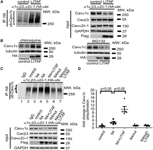
LITAF (lipopolysaccharide-induced tumor necrosis factor)-mediated ubiquitination and degradation of Cavα1c (L-type calcium channel alpha-1C subunit) in tsA201 cells. Cells were transfected with plasmids for Cavα1c, Cavβ3, and Cavα2δ-1, HA (hemagglutinin)-tagged ubiquitin (HA-ubi), GFP (green fluorescence protein) as control, or Flag-tagged LITAF. Immunoprecipitation (IP) of lysates from transfected cells was performed with anti-HA antibody. A, A representative immunoblot shows levels of ubiquitinated Cavα1c ( left) and input levels of Cavα1c, Cavβ3, Cavα2δ-1, Flag-tagged LITAF, and GAPDH ( right). B, LITAF-mediated degradation of Cavα1c through lysosomes. Cells were transfected with plasmids for Cavα1c, Cavβ3, and Cavα2δ-1, GFP as control, or HA-tagged LITAF for 24 h and then treated with 10 µM chloroquine or 5 µM MG132 (N-benzyloxycarbonyl-L-leucyl-L-leucyl-L-leucinal) for 20 h. Representative Western blots show total abundance of Cavα1c and tubulin of treated cells. C, IP of lysates from cells transfected with plasmids for Cavα1c, Cavβ3, and Cavα2δ-1, HA-tagged ubiquitin, GFP as control, NEDD (neural precursor cell expressed developmentally down-regulated protein) 4-1 (N4), NEDD4-1-C867A (N4mut), or Flag-tagged LITAF was performed with anti-HA antiserum. A representative immunoblot shows levels of ubiquitinated Cavα1c ( top) and input levels of Cavα1c, Cavβ3, Cavα2δ-1, Flag-tagged LITAF, and GAPDH ( bottom). D, Respective changes in the level of ubiquitinated Cavα1c, normalized to total Cavα1c (5 experiments, performed in duplicate; mean±SEM). Student t test, P<0.05. |