
Fig. 4
- ID
- ZDB-FIG-240509-87
- Publication
- Moshal et al., 2019 - LITAF Regulates Cardiac L-type Calcium Channels by Modulating NEDD4-1 Ubiquitin Ligase
- Other Figures
- All Figure Page
- Back to All Figure Page
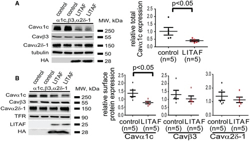
Functional interaction between LITAF (lipopolysaccharide-induced tumor necrosis factor) and L-type calcium channel (LTCC) in tsA201 cells. Cells were transfected with plasmids for Cavα1c, (L-type calcium channel alpha-1C subunit) Cavβ3, and Cavα2δ-1 to reconstitute functional LTCC, GFP (green fluorescence protein), or HA (hemagglutinin)-tagged LITAF. Cell-surface protein levels were determined by biotinylation: cell-surface proteins were biotinylated using sulfo-NHS-SS-biotin, purified with neutravidin beads from total cell lysates, subjected to SDS-PAGE and blotted onto a nitrocellulose membrane. A, A representative Western blot shows total protein levels of Cavα1c, Cavβ3, Cavα2δ-1, HA-LITAF, and tubulin ( left). Respective change in total Cavα1c abundance, normalized to tubulin levels (n=5, performed in triplicate; mean±SEM). Student t test, P<0.05 ( right). B, A representative Western blot shows cell-surface protein levels of Cavα1c, Cavβ3, Cavα2δ-1, TFR (transferrin receptor), total LITAF, and HA-LITAF ( left). Respective changes in cell membrane protein levels of Cavα1c, Cavβ3, and Cavα2δ-1 normalized to transferrin receptor levels (n=5, performed in triplicate; mean±SEM). Student t test, P<0.05 ( right). |