
Fig. 2
- ID
- ZDB-FIG-240426-18
- Publication
- Tran et al., 2024 - Caffeine-induced protein kinase A activation restores cognitive deficits induced by sleep deprivation by regulating O-GlcNAc cycling in adult zebrafish
- Other Figures
- All Figure Page
- Back to All Figure Page
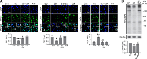
Effects of caffeine on brain O-GlcNAcylation in brain of sleep-deprived zebrafish. Sleep deprivation (SD) was induced for 3 days with or without daily injections of caffeine (10 μg/g) for 3 days. A: representative IF staining images (×40) and quantification graphs of anti-O-GlcNAc, OGT, and OGA are presented. O-GlcNAc (n = 6 fish/group), OGT (n = 3 fish/group), and OGA (n = 3 fish/group) were stained in green, whereas nuclei were counterstained with DAPI (blue) (*P < 0.05, **P < 0.01, ***P < 0.001 vs. Con; #P < 0.05, ##P < 0.01 vs. SD). Enlarged images are shown in the white box. Scale bar = 20 μm. B: representative Western blot images and the quantification graph of O-GlcNAc and β-actin of Con, SD, and SD+Caf are depicted from protein lysate extracted from zebrafish brains (n = 5 replicates/group, *P < 0.05 vs. Control; #P < 0.05 vs. SD). The Kruskal–Wallis test followed by FDR correction was used to assess the statistical significance of immunofluorescence staining and Western blot quantification. Caf, caffeine; Con, control; FDR, false discovery rate; IF, immunofluorescence; OGA, O-GlcNAcase; OGT, O-GlcNAc transferase. |