
Fig. 3
- ID
- ZDB-FIG-240419-73
- Publication
- Yao et al., 2024 - Rab11 promotes single Mauthner cell axon regeneration in vivo through axon guidance molecule Ntng2b
- Other Figures
- All Figure Page
- Back to All Figure Page
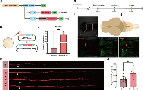
Rab11bb overexpression promoted the capacity of M-cells axon regeneration. (A) Plasmid construction legend of the control group and Rab11bb overexpression plasmids. The CMV-GAL4-VP16 plasmid drives the expression of UAS-mCherry and UAS-rab11bb-mCherry plasmids (CMV: Cytomegalovirus; GAL4: Galectin-4; PA: poly adenylate). (B) Schematic diagram of microinjection using the two-plasmid system. (C) qRT-PCR analysis of rab11bb overexpression efficiency. The total RNA of each group (n = 30) was obtained from three independent experiments. Assessed by unpaired, two-tailed Student's t-test. *** p < 0.001. (D) Timeline of time points of electroporation, axotomy and imaging. (E) Pattern Diagram of electroporation and confocal images of positive expression in M-cell. The white box represents the position of M-cell under 40 × magnification in 10 × magnification field. Scale bar, 50 μm. (EGFP: labeled M-cells in Tol-056 zebrafish strain; mCherry: fluorescent reporter gene in foreign plasmid). (F) Representative diagram of confocal imaging of M-cells axon regeneration. Asterisk, ablation site; Scale bar, 50 μm. (Ctrl: control; OE: overexpression). (G) Statistical quantitative diagram of axon regeneration (control: 273.0 ± 31.71 μm, n = 10; Rab11bb overexpression: 470.5 ± 43.82 μm, n = 9). Assessed by unpaired, two-tailed Student's t-test. ** p < 0.01. |