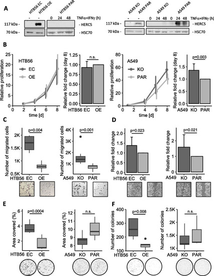
HERC5 inhibits tumor-associated aggressiveness in vitro. A Western Blot showing the HERC5 protein levels in the HTB56 OE/EC and A549 PAR/KO model cell lines with and without stimulation. B HTB56 EC/OE showed no significant differences in proliferation as assessed by a Methylene Blue proliferation assay (p = 0.438, n = 6, one sample t-test), while A549 KO/PAR cells showed a significant increase in proliferation at low HERC5 levels (p = 0.003, n = 11, one sample t-test). C HTB56 EC and A549 KO cells have an increased migratory potential compared to HTB56 OE (p = 0.004, n = 4, Student’s t-test) and A549 PAR cells (p = 0.001, n = 7, Mann–Whitney U test), respectively. D The wound healing assay revealed a higher ratio of migration toward the scratch in HTB56 EC compared to HERC5 OE (p = 0.023, n = 4, one sample t-test) and A549 HERC5 KO compared to PAR cells (p = 0.021, n = 3, one sample t-test) after 18 h. E and F Overexpression of HERC5 caused a decrease in clonogenic and anchorage-independent growth (p = 0.0004, n = 5, Student’s t-test; p = 0.008, n = 5, Student’s t-test, respectively) but not in A549 cells (p = 0.069, Student’s t-test, n = 5; p = 0.515, Student’s t-test, n = 5, respectively). Error bars in bar plots are depicting the 95% confidence interval
|
