FIGURE

Fig. 2.

ID
ZDB-FIG-240408-45
Publication
Chen et al., 2024 - col1a2+ fibroblasts/muscle progenitors finetune xanthophore countershading by differentially expressing csf1a/1b in embryonic zebrafish
Other Figures
All Figure Page
Back to All Figure Page
Fig. 2.

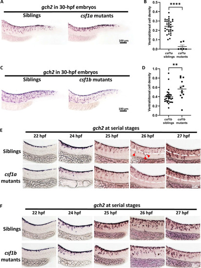
Xanthophore countershading is abnormal in csf1a and csf1b mutants.

(A and B) Representative images (A) and quantification (B) of xanthophores in csf1a siblings (n = 32) and mutants (n = 6). Xanthophores are indicated by WISH staining of gch2. Unpaired Student’s t tests were used to calculate the P value. Two-tailed P values are used. Error bars represent means ± SD. ****P < 0.0001. (C and D) Representative images (C) and quantification (D) of xanthophores in csf1b siblings (n = 30) and mutants (n = 12). Xanthophores are indicated by WISH staining of gch2. Unpaired Student’s t tests were used to calculate the P value. Two-tailed P values are used. Error bars represent means ± SD. ns, P > 0.05; **P < 0.01. (E) WISH staining at serial stages shows that xanthoblasts in csf1a mutants fail to cross the horizontal myoseptum at about 26 hpf. The horizontal myoseptum is indicated by the white dotted line. (F) WISH staining at serial stages shows that xanthoblasts in csf1b mutants migrate normally toward the ventrum.

Expression Data

Expression Detail
Antibody Labeling
Phenotype Data

Phenotype Detail
Acknowledgments
This image is the copyrighted work of the attributed author or publisher, and ZFIN has permission only to display this image to its users. Additional permissions should be obtained from the applicable author or publisher of the image. Full text @ Sci Adv