Fig. 1
- ID
- ZDB-FIG-240405-8
- Publication
- Salgado-Almario et al., 2023 - ERG potassium channels and T-type calcium channels contribute to the pacemaker and atrioventricular conduction in zebrafish larvae
- Other Figures
- All Figure Page
- Back to All Figure Page
|
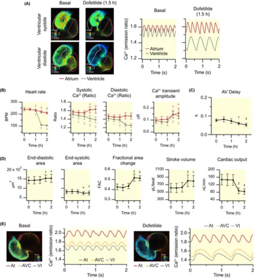
Effect of dofetilide on cardiac Ca2+ levels and ventricular contractility of 3 dpf zebrafish larvae. Dofetilide (100 μM) was added to Tg(myl7:Twitch-4) larvae at 3 dpf (N = 3, n = 9) after recording basal Ca2+ levels. (A) Emission ratio images of a representative larva before (basal) and after 1.5 h incubation with dofetilide. The calibration squares show the distance in μm (top side), the emission ratio coded in the hue (right side), and the fluorescence intensity (bottom side). The traces show the atrial (red) and ventricular (gray) Ca2+ levels (emission ratio). (B) Effect of dofetilide on heart rate, systolic and diastolic Ca2+ levels, and Ca2+ transient amplitude in the atrium (red) and ventricle (gray). (C) Effect of dofetilide on the AV delay (time between the start of atrial and ventricular Ca2+ transients). (D) Effect of dofetilide on ventricular areas, fractional area change, stroke volume, and cardiac output. (E) Effect of dofetilide on AV conduction in a 3 dpf zebrafish larva whose heart was stopped by incubation with the myosin inhibitor para-aminoblebbistatin (75 μM for 2 h) (representative experiment). ROIs were drawn as indicated to investigate the progression of the Ca2+ wave from the atrium (At), across the AV canal (AVC), and into the ventricle (Vt). The traces show the time course of the Ca2+ transients in each ROI. Data in B, C, and D are shown as the mean ± SD. Statistical analysis was performed using a one-way anova test with Dunnett's multiple comparisons post-test (*p < 0.05, †p < 0.01, ‡p < 0.001) |

