|
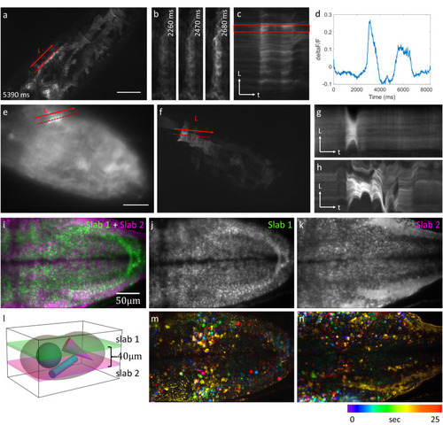
Selective projection imaging of calcium dynamics. a–d Calcium dynamics in a Drosophila embryo labeled with jGCaMP7s-CAAX imaged at a rate of 50 Hz using OPMprops. b Zoomed-in views of the area surrounding the dashed line in (a). c Kymograph along the dotted line in (a). d Calcium signal analysis (dF/F) from the box in (c). e Projection image of another jGCaMP7s-CAAX labeled embryo using OPMpro. f Same embryo as in (e), but imaged with OPMprops at 20 Hz framerate. g Kymograph along the dotted line in (e). h Kymograph along the dotted line in (f). i–n OPMprops imaging at two depths within a zebrafish larva labeled with Tg(elavl3:soma-GCaMP7f). i Overlay of the two projections. j, k Individual projections. l Schematic representation of the projection imaging. The two sub-volumes (“slabs”) are separated by 40 microns. m–n Color-coded timeseries corresponding to j, k where the static background was subtracted. Both the Drosophila embryo and zebrafish larva imaging were repeated independently 3 times with similar results. Source data are provided as a Source Data file.
|